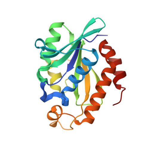
Structural and binding studies of peptidyl-tRNA hydrolase from Pseudomonas aeruginosa provide a platform for the structure-based inhibitor design against peptidyl-tRNA hydrolase
Singh, A., Kumar, A., Gautam, L., Sharma, P., Sinha, M., Bhushan, A., Kaur, P., Sharma, S., Arora, A., Singh, T.P.(2014) Biochem J 463: 329-337
- PubMed: 25101795
- DOI: https://doi.org/10.1042/BJ20140631
- Primary Citation of Related Structures:
4FNO, 4JC4, 4QAJ, 4QBK, 4QD3 - PubMed Abstract:
During the course of protein synthesis in the cell, the translation process is often terminated due to various reasons. As a result, peptidyl-tRNA molecules are released which are toxic to the cell as well reducing the availability of free amino acid and tRNA molecules for the required protein synthesis in the cell. Such a situation is corrected by an enzyme, Pth (peptidyl-tRNA hydrolase), which catalyses the release of free tRNA and peptide moieties from peptidyl-tRNAs. This means that the active Pth is essential for the survival of bacteria. In order to design inhibitors of PaPth (Pth from Pseudomonas aeruginosa), we determined the structures of PaPth in its native and bound states with compounds amino acylate-tRNA analogue and 5-azacytidine. The structure determination of the native protein revealed that the substrate-binding site was partially occupied by Glu161 from the neigh-bouring molecule. The structure of PaPth indicated that the substrate-binding site can be broadly divided into three distinct subsites. The structures of the two complexes showed that the amino acylate-tRNA analogue filled three subsites, whereas 5-azacytidine filled two subsites. The common sugar and the base moieties of the two compounds occupied identical positions in the cleft. Using surface plasmon resonance, the dissociation constants for the amino acylate-tRNA analogue and 5-azacytidine were found to be 3.53×10⁻⁸ M and 5.82×10⁻⁸ M respectively.
Organizational Affiliation:
*Department of Biophysics, All India Institute of Medical Sciences, Ansari Nagar, New Delhi, 110029 Delhi, India.