4F1H

Crystal structure of TDP2 from Danio rerio complexed with a single strand DNA


Experimental Data Snapshot

  • Method: X-RAY DIFFRACTION
  • Resolution: 1.66 Å
  • R-Value Free: 0.193 
  • R-Value Work: 0.164 
  • R-Value Observed: 0.164 

wwPDB Validation   3D Report Full Report


This is version 1.2 of the entry. See complete history


Literature

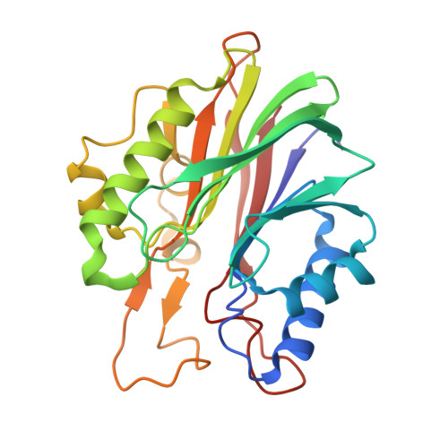
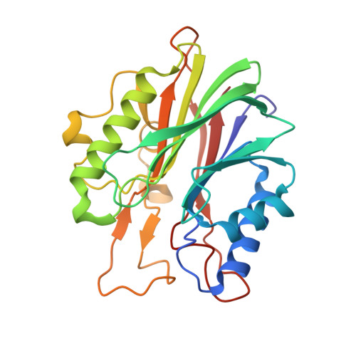
Structural basis for recognition of 5'-phosphotyrosine adducts by Tdp2.

Shi, K.Kurahashi, K.Gao, R.Tsutakawa, S.E.Tainer, J.A.Pommier, Y.Aihara, H.

(2012) Nat Struct Mol Biol 19: 1372-1377

  • DOI: https://doi.org/10.1038/nsmb.2423
  • Primary Citation of Related Structures:  
    4F1H, 4F1I, 4FPV, 4FVA, 4GEW

  • PubMed Abstract: 

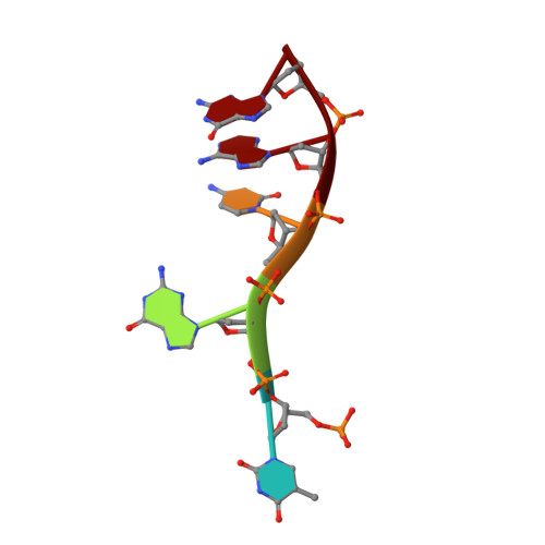
    The DNA-repair enzyme Tdp2 resolves 5'-phosphotyrosyl DNA adducts and mediates resistance to anticancer drugs that target covalent topoisomerase-DNA complexes. Tdp2 also participates in key signaling pathways during development and tumorigenesis and cleaves a protein-RNA linkage during picornavirus replication. The crystal structure of zebrafish Tdp2 bound to DNA reveals a deep, narrow basic groove that selectively accommodates the 5' end of single-stranded DNA in a stretched conformation. The crystal structure of the full-length Caenorhabditis elegans Tdp2 shows that this groove can also accommodate an acidic peptide stretch in vitro, with glutamate and aspartate side chains occupying the DNA backbone phosphate-binding sites. This extensive molecular mimicry suggests a potential mechanism for autoregulation and interaction of Tdp2 with phosphorylated proteins in signaling. Our study provides a framework to interrogate functions of Tdp2 and develop inhibitors for chemotherapeutic and antiviral applications.


  • Organizational Affiliation

    Department of Biochemistry, Molecular Biology and Biophysics, University of Minnesota, Minneapolis, Minnesota, USA.


Macromolecules

Find similar proteins by:  (by identity cutoff)  |  3D Structure
Entity ID: 2
MoleculeChains Sequence LengthOrganismDetailsImage
Tyrosyl-DNA phosphodiesterase 2B [auth A]250Danio rerioMutation(s): 0 
Gene Names: tdp2ttrapttraplsi:ch211-81e5.5si:dkey-218n20.5
EC: 3.1.4
UniProt
Find proteins for Q5XJA0 (Danio rerio)
Explore Q5XJA0 
Go to UniProtKB:  Q5XJA0
Entity Groups  
Sequence Clusters30% Identity50% Identity70% Identity90% Identity95% Identity100% Identity
UniProt GroupQ5XJA0
Sequence Annotations
Expand
  • Reference Sequence
Find similar proteins by:  (by identity cutoff)  |  3D Structure
Entity ID: 3
MoleculeChains Sequence LengthOrganismDetailsImage
Tyrosyl-DNA phosphodiesterase 2C [auth B]251Danio rerioMutation(s): 0 
Gene Names: tdp2ttrapttraplsi:ch211-81e5.5si:dkey-218n20.5
EC: 3.1.4
UniProt
Find proteins for Q5XJA0 (Danio rerio)
Explore Q5XJA0 
Go to UniProtKB:  Q5XJA0
Entity Groups  
Sequence Clusters30% Identity50% Identity70% Identity90% Identity95% Identity100% Identity
UniProt GroupQ5XJA0
Sequence Annotations
Expand
  • Reference Sequence

Find similar nucleic acids by:  Sequence   |   3D Structure  

Entity ID: 1
MoleculeChains LengthOrganismImage
DNA (5'-D(P*TP*GP*CP*AP*G)-3')A [auth C]5N/A
Sequence Annotations
Expand
  • Reference Sequence
Experimental Data & Validation

Experimental Data

  • Method: X-RAY DIFFRACTION
  • Resolution: 1.66 Å
  • R-Value Free: 0.193 
  • R-Value Work: 0.164 
  • R-Value Observed: 0.164 
  • Space Group: P 21 21 21
Unit Cell:
Length ( Å )Angle ( ˚ )
a = 57.918α = 90
b = 95.1β = 90
c = 104.732γ = 90
Software Package:
Software NamePurpose
ADSCdata collection
PHASERphasing
PHENIXrefinement
HKL-2000data reduction
HKL-2000data scaling

Structure Validation

View Full Validation Report


Entry History 

Deposition Data

Revision History  (Full details and data files)

  • Version 1.0: 2012-10-31
    Type: Initial release
  • Version 1.1: 2013-01-02
    Changes: Database references
  • Version 1.2: 2024-02-28
    Changes: Data collection, Database references, Derived calculations