4EUL

Crystal structure of enhanced Green Fluorescent Protein to 1.35A resolution reveals alternative conformations for Glu222


Experimental Data Snapshot

  • Method: X-RAY DIFFRACTION
  • Resolution: 1.35 Å
  • R-Value Free: 0.169 
  • R-Value Work: 0.128 
  • R-Value Observed: 0.130 

wwPDB Validation   3D Report Full Report


This is version 1.3 of the entry. See complete history


Literature

Crystal structure of enhanced green fluorescent protein to 1.35 a resolution reveals alternative conformations for glu222.

Arpino, J.A.Rizkallah, P.J.Jones, D.D.

(2012) PLoS One 7: e47132-e47132

  • DOI: https://doi.org/10.1371/journal.pone.0047132
  • Primary Citation of Related Structures:  
    4EUL

  • PubMed Abstract: 

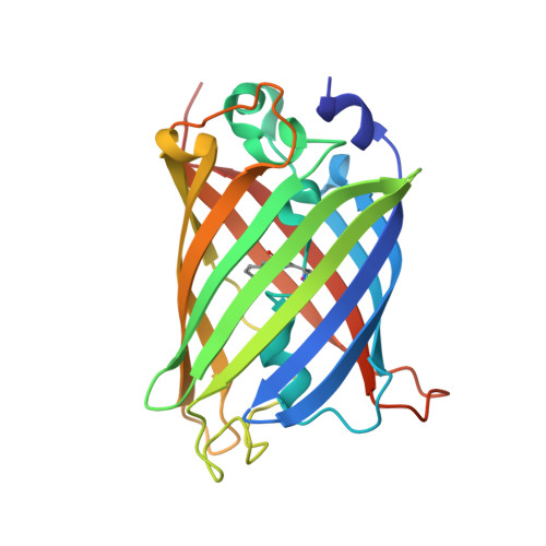
    Enhanced Green Fluorescent Protein (EGFP) is one of the most widely used engineered variants of the original wild-type Green Fluorescent Protein. Here, we report the high resolution (1.35 Å) structure of EGFP crystallised in its untagged sequence form that reveals the combined impact of the F64L and S65T, that give rise to improved folding and spectral characteristics. The overall structure of EGFP is very similar to wt GFP, forming the classical β-barrel fold with the chromophore containing helix running through the core of the structure. Replacement of Phe64 with Leu in EGFP results in subtle rearrangement of hydrophobic core packing close to the chromophore including the reduction in surface exposure of two hydrophobic residues. Replacement of Ser65 with Thr has a significant impact on the local hydrogen bond network in the vicinity of the chromophore. Detailed analysis of electron density reveals that several residues close to the chromophore occupy at least two distinct conformations. This includes Glu222 that defines the charged state on the chromophore, with the two conformations having slightly different effects on the hydrogen bond network surrounding the chromophore. Hence, the reported high-resolution structure of EGFP has provided a long overdue molecular description of one of the most important fluorescent protein variants currently in general use.


  • Organizational Affiliation

    School of Biosciences, Cardiff University, Cardiff, United Kingdom.


Macromolecules
Find similar proteins by:  (by identity cutoff)  |  3D Structure
Entity ID: 1
MoleculeChains Sequence LengthOrganismDetailsImage
Green fluorescent protein237Aequorea victoriaMutation(s): 2 
Gene Names: GFP
UniProt
Find proteins for P42212 (Aequorea victoria)
Explore P42212 
Go to UniProtKB:  P42212
Entity Groups  
Sequence Clusters30% Identity50% Identity70% Identity90% Identity95% Identity100% Identity
UniProt GroupP42212
Sequence Annotations
Expand
  • Reference Sequence
Small Molecules
Modified Residues  1 Unique
IDChains TypeFormula2D DiagramParent
CRO
Query on CRO
A
L-PEPTIDE LINKINGC15 H17 N3 O5THR, TYR, GLY
Experimental Data & Validation

Experimental Data

  • Method: X-RAY DIFFRACTION
  • Resolution: 1.35 Å
  • R-Value Free: 0.169 
  • R-Value Work: 0.128 
  • R-Value Observed: 0.130 
  • Space Group: P 21 21 21
Unit Cell:
Length ( Å )Angle ( ˚ )
a = 51.114α = 90
b = 62.246β = 90
c = 69.641γ = 90
Software Package:
Software NamePurpose
xia2data scaling
MOLREPphasing
REFMACrefinement
XDSdata reduction
SCALAdata scaling

Structure Validation

View Full Validation Report


Entry History 

Deposition Data

Revision History  (Full details and data files)

  • Version 1.0: 2012-10-03
    Type: Initial release
  • Version 1.1: 2012-12-19
    Changes: Database references
  • Version 1.2: 2023-09-13
    Changes: Data collection, Database references, Derived calculations, Refinement description
  • Version 1.3: 2023-12-06
    Changes: Data collection