4ESP

Crystal Structure of Peanut Allergen Ara h 5


Experimental Data Snapshot

  • Method: X-RAY DIFFRACTION
  • Resolution: 1.10 Å
  • R-Value Free: 0.183 
  • R-Value Work: 0.168 
  • R-Value Observed: 0.169 

wwPDB Validation   3D Report Full Report


This is version 1.2 of the entry. See complete history


Literature

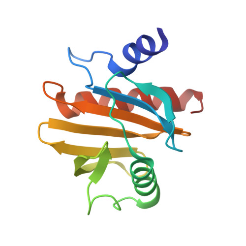
Crystal Structure of Peanut ( Arachis hypogaea ) Allergen Ara h 5.

Wang, Y.Fu, T.J.Howard, A.Kothary, M.H.McHugh, T.H.Zhang, Y.

(2013) J Agric Food Chem 61: 1573-1578

  • DOI: https://doi.org/10.1021/jf303861p
  • Primary Citation of Related Structures:  
    4ESP

  • PubMed Abstract: 

    Profilins from numerous species are known to be allergens, including food allergens, such as peanut ( Arachis hypogaea ) allergen Ara h 5, and pollen allergens, such as birch allergen Bet v 2. Patients with pollen allergy can also cross-react to peanut. Structural characterization of allergens will allow a better understanding of the allergenicity of food allergens and their cross-reactivities. The three-dimensional structures of most known food allergens remain to be elucidated. Here, we report the first crystallographic study of a food allergen in the profilin family. The structure of peanut allergen Ara h 5 was determined, and the resolution of the final refined structure was 1.1 Å. Structure alignment revealed that Ara h 5 is more similar to Bet v 2 than to Hev b 8, although sequence alignment suggested that Ara h 5 is more closely related to Hev b 8 than to Bet v 2, indicating that homology-model-based prediction of immunoglobulin E epitopes needs to be interpreted with caution.


  • Organizational Affiliation

    Department of Biological and Chemical Sciences, Illinois Institute of Technology, Chicago, Illinois 60616, United States.


Macromolecules
Find similar proteins by:  (by identity cutoff)  |  3D Structure
Entity ID: 1
MoleculeChains Sequence LengthOrganismDetailsImage
Profilin130Arachis hypogaeaMutation(s): 0 
Gene Names: profilin
UniProt
Find proteins for D3K177 (Arachis hypogaea)
Explore D3K177 
Go to UniProtKB:  D3K177
Entity Groups  
Sequence Clusters30% Identity50% Identity70% Identity90% Identity95% Identity100% Identity
UniProt GroupD3K177
Sequence Annotations
Expand
  • Reference Sequence
Small Molecules
Ligands 2 Unique
IDChains Name / Formula / InChI Key2D Diagram3D Interactions
EDO
Query on EDO

Download Ideal Coordinates CCD File 
B [auth A]
C [auth A]
D [auth A]
E [auth A]
F [auth A]
B [auth A],
C [auth A],
D [auth A],
E [auth A],
F [auth A],
G [auth A],
H [auth A],
I [auth A]
1,2-ETHANEDIOL
C2 H6 O2
LYCAIKOWRPUZTN-UHFFFAOYSA-N
IPA
Query on IPA

Download Ideal Coordinates CCD File 
J [auth A],
K [auth A]
ISOPROPYL ALCOHOL
C3 H8 O
KFZMGEQAYNKOFK-UHFFFAOYSA-N
Experimental Data & Validation

Experimental Data

  • Method: X-RAY DIFFRACTION
  • Resolution: 1.10 Å
  • R-Value Free: 0.183 
  • R-Value Work: 0.168 
  • R-Value Observed: 0.169 
  • Space Group: P 41 21 2
Unit Cell:
Length ( Å )Angle ( ˚ )
a = 58.574α = 90
b = 58.574β = 90
c = 101.717γ = 90
Software Package:
Software NamePurpose
DENZOdata reduction
SCALEPACKdata scaling
PHASERphasing
REFMACrefinement
PDB_EXTRACTdata extraction

Structure Validation

View Full Validation Report


Entry History 

Deposition Data

Revision History  (Full details and data files)

  • Version 1.0: 2013-03-06
    Type: Initial release
  • Version 1.1: 2017-11-15
    Changes: Refinement description
  • Version 1.2: 2024-02-28
    Changes: Data collection, Database references, Derived calculations