4EL9

Structure of N-terminal kinase domain of RSK2 with afzelin


Experimental Data Snapshot

  • Method: X-RAY DIFFRACTION
  • Resolution: 1.55 Å
  • R-Value Free: 0.226 
  • R-Value Work: 0.200 
  • R-Value Observed: 0.226 

wwPDB Validation   3D Report Full Report


Ligand Structure Quality Assessment 


This is version 1.1 of the entry. See complete history


Literature

Insights into the Inhibition of the p90 Ribosomal S6 Kinase (RSK) by the Flavonol Glycoside SL0101 from the 1.5 A Crystal Structure of the N-Terminal Domain of RSK2 with Bound Inhibitor.

Utepbergenov, D.Derewenda, U.Olekhnovich, N.Szukalska, G.Banerjee, B.Hilinski, M.K.Lannigan, D.A.Stukenberg, P.T.Derewenda, Z.S.

(2012) Biochemistry 51: 6499-6510

  • DOI: https://doi.org/10.1021/bi300620c
  • Primary Citation of Related Structures:  
    3UBD, 4EL9

  • PubMed Abstract: 

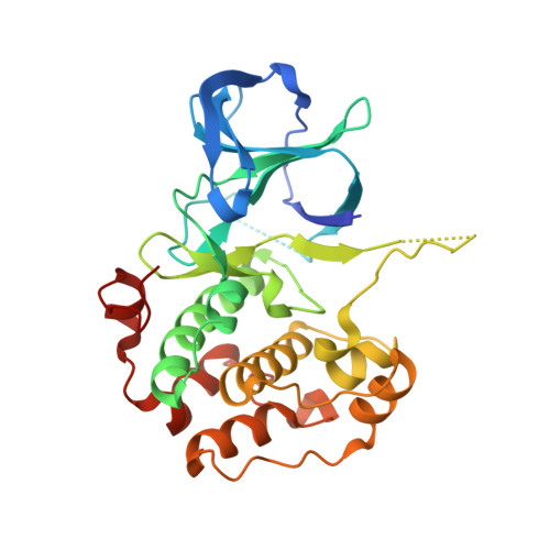
    The p90 ribosomal S6 family of kinases (RSK) are potential drug targets, due to their involvement in cancer and other pathologies. There are currently only two known selective inhibitors of RSK, but the basis for selectivity is not known. One of these inhibitors is a naturally occurring kaempferol-α-L-diacetylrhamnoside, SL0101. Here, we report the crystal structure of the complex of the N-terminal kinase domain of the RSK2 isoform with SL0101 at 1.5 Å resolution. The refined atomic model reveals unprecedented structural reorganization of the protein moiety, as compared to the nucleotide-bound form. The entire N-lobe, the hinge region, and the αD-helix undergo dramatic conformational changes resulting in a rearrangement of the nucleotide binding site with concomitant formation of a highly hydrophobic pocket spatially suited to accommodate SL0101. These unexpected results will be invaluable in further optimization of the SL0101 scaffold as a promising lead for a novel class of kinase inhibitors.


  • Organizational Affiliation

    Department of Molecular Physiology and Biological Physics, University of Virginia, School of Medicine, Charlottesville, VA 22908, USA.


Macromolecules
Find similar proteins by:  (by identity cutoff)  |  3D Structure
Entity ID: 1
MoleculeChains Sequence LengthOrganismDetailsImage
Ribosomal protein S6 kinase alpha-3305Mus musculusMutation(s): 0 
Gene Names: Rps6ka3Mapkapk1bRps6ka-rs1Rsk2
EC: 2.7.11.1
UniProt
Find proteins for P18654 (Mus musculus)
Explore P18654 
Go to UniProtKB:  P18654
Entity Groups  
Sequence Clusters30% Identity50% Identity70% Identity90% Identity95% Identity100% Identity
UniProt GroupP18654
Sequence Annotations
Expand
  • Reference Sequence
Small Molecules
Ligands 1 Unique
IDChains Name / Formula / InChI Key2D Diagram3D Interactions
AFE
Query on AFE

Download Ideal Coordinates CCD File 
B [auth A]5,7-dihydroxy-2-(4-hydroxyphenyl)-4-oxo-4H-chromen-3-yl 6-deoxy-alpha-L-mannopyranoside
C21 H20 O10
SOSLMHZOJATCCP-AEIZVZFYSA-N
Binding Affinity Annotations 
IDSourceBinding Affinity
AFE PDBBind:  4EL9 IC50: 4370 (nM) from 1 assay(s)
Experimental Data & Validation

Experimental Data

  • Method: X-RAY DIFFRACTION
  • Resolution: 1.55 Å
  • R-Value Free: 0.226 
  • R-Value Work: 0.200 
  • R-Value Observed: 0.226 
  • Space Group: C 1 2 1
Unit Cell:
Length ( Å )Angle ( ˚ )
a = 99.119α = 90
b = 40.86β = 114.64
c = 83.867γ = 90
Software Package:
Software NamePurpose
SERGUIdata collection
AMoREphasing
PHENIXrefinement
HKL-2000data reduction
HKL-2000data scaling

Structure Validation

View Full Validation Report


Ligand Structure Quality Assessment 


Entry History 

Deposition Data

Revision History  (Full details and data files)

  • Version 1.0: 2012-09-05
    Type: Initial release
  • Version 1.1: 2023-09-13
    Changes: Data collection, Database references, Derived calculations, Refinement description, Structure summary