4EIK

Crystal Structure of the Human Fyn SH3 domain in complex with the synthetic peptide VSL12


Experimental Data Snapshot

  • Method: X-RAY DIFFRACTION
  • Resolution: 1.60 Å
  • R-Value Free: 0.195 
  • R-Value Work: 0.174 
  • R-Value Observed: 0.175 

wwPDB Validation   3D Report Full Report


This is version 1.4 of the entry. See complete history


Literature

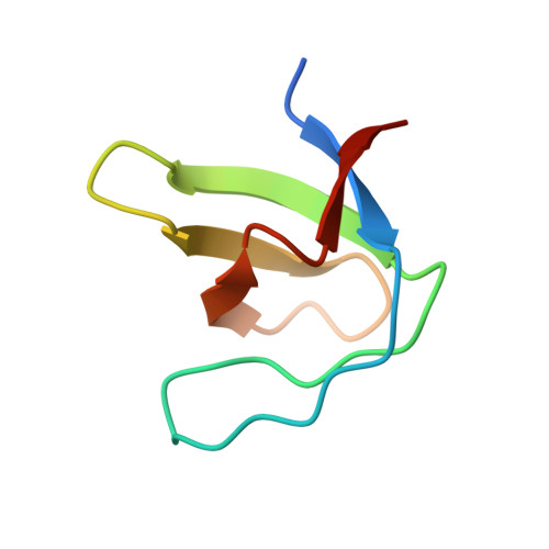
Crystallization of and selenomethionine phasing strategy for a SETMAR-DNA complex.

Chen, Q.Georgiadis, M.

(2016) Acta Crystallogr F Struct Biol Commun 72: 713-719

  • DOI: https://doi.org/10.1107/S2053230X16012723
  • Primary Citation of Related Structures:  
    4EIK

  • PubMed Abstract: 

    Transposable elements have played a critical role in the creation of new genes in all higher eukaryotes, including humans. Although the chimeric fusion protein SETMAR is no longer active as a transposase, it contains both the DNA-binding domain (DBD) and catalytic domain of the Hsmar1 transposase. The amino-acid sequence of the DBD has been virtually unchanged in 50 million years and, as a consequence, SETMAR retains its sequence-specific binding to the ancestral Hsmar1 terminal inverted repeat (TIR) sequence. Thus, the DNA-binding activity of SETMAR is likely to have an important biological function. To determine the structural basis for the recognition of TIR DNA by SETMAR, the design of TIR-containing oligonucleotides and SETMAR DBD variants, crystallization of DBD-DNA complexes, phasing strategies and initial phasing experiments are reported here. An unexpected finding was that oligonucleotides containing two BrdUs in place of thymidines produced better quality crystals in complex with SETMAR than their natural counterparts.


  • Organizational Affiliation

    Biochemistry and Molecular Biology, Indiana University School of Medicine, 635 Barnhill Drive, Indianapolis, IN 46202, USA.


Macromolecules
Find similar proteins by:  (by identity cutoff)  |  3D Structure
Entity ID: 1
MoleculeChains Sequence LengthOrganismDetailsImage
Tyrosine-protein kinase Fyn64Homo sapiensMutation(s): 0 
EC: 2.7.10.2
UniProt & NIH Common Fund Data Resources
Find proteins for P06241 (Homo sapiens)
Explore P06241 
Go to UniProtKB:  P06241
PHAROS:  P06241
GTEx:  ENSG00000010810 
Entity Groups  
Sequence Clusters30% Identity50% Identity70% Identity90% Identity95% Identity100% Identity
UniProt GroupP06241
Sequence Annotations
Expand
  • Reference Sequence

Find similar proteins by:  Sequence   |   3D Structure  

Entity ID: 2
MoleculeChains Sequence LengthOrganismDetailsImage
VSL12 peptide12synthetic constructMutation(s): 0 
Entity Groups  
Sequence Clusters30% Identity50% Identity70% Identity90% Identity95% Identity100% Identity
Sequence Annotations
Expand
  • Reference Sequence
Small Molecules
Ligands 1 Unique
IDChains Name / Formula / InChI Key2D Diagram3D Interactions
NA
Query on NA

Download Ideal Coordinates CCD File 
C [auth A]SODIUM ION
Na
FKNQFGJONOIPTF-UHFFFAOYSA-N
Experimental Data & Validation

Experimental Data

  • Method: X-RAY DIFFRACTION
  • Resolution: 1.60 Å
  • R-Value Free: 0.195 
  • R-Value Work: 0.174 
  • R-Value Observed: 0.175 
  • Space Group: P 63
Unit Cell:
Length ( Å )Angle ( ˚ )
a = 81.843α = 90
b = 81.843β = 90
c = 35.904γ = 120
Software Package:
Software NamePurpose
SCALAdata scaling
MOLREPphasing
REFMACrefinement
PDB_EXTRACTdata extraction
ADSCdata collection
DENZOdata reduction
SCALEPACKdata scaling

Structure Validation

View Full Validation Report


Entry History 

Deposition Data

Revision History  (Full details and data files)

  • Version 1.0: 2013-04-10
    Type: Initial release
  • Version 1.1: 2016-09-14
    Changes: Database references
  • Version 1.2: 2017-11-15
    Changes: Refinement description
  • Version 1.3: 2018-04-18
    Changes: Data collection, Database references
  • Version 1.4: 2023-09-13
    Changes: Data collection, Database references, Derived calculations, Refinement description