4EHS

Crystal structure of Helicobacter pylori DnaG Primase C terminal domain


Experimental Data Snapshot

  • Method: X-RAY DIFFRACTION
  • Resolution: 1.78 Å
  • R-Value Free: 0.249 
  • R-Value Work: 0.195 
  • R-Value Observed: 0.198 

wwPDB Validation   3D Report Full Report


This is version 1.3 of the entry. See complete history


Literature

Crystal structure and mode of helicase binding of the C-terminal domain of primase from Helicobacter pylori

Abdul Rehman, S.A.Verma, V.Mazumder, M.Dhar, S.K.Gourinath, S.

(2013) J Bacteriol 195: 2826-2838

  • DOI: https://doi.org/10.1128/JB.00091-13
  • Primary Citation of Related Structures:  
    4EHS

  • PubMed Abstract: 

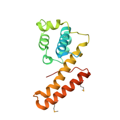
    To better understand the poor conservation of the helicase binding domain of primases (DnaGs) among the eubacteria, we determined the crystal structure of the Helicobacter pylori DnaG C-terminal domain (HpDnaG-CTD) at 1.78 Å. The structure has a globular subdomain connected to a helical hairpin. Structural comparison has revealed that globular subdomains, despite the variation in number of helices, have broadly similar arrangements across the species, whereas helical hairpins show different orientations. Further, to study the helicase-primase interaction in H. pylori, a complex was modeled using the HpDnaG-CTD and HpDnaB-NTD (helicase) crystal structures using the Bacillus stearothermophilus BstDnaB-BstDnaG-CTD (helicase-primase) complex structure as a template. By using this model, a nonconserved critical residue Phe534 on helicase binding interface of DnaG-CTD was identified. Mutation guided by molecular dynamics, biophysical, and biochemical studies validated our model. We further concluded that species-specific helicase-primase interactions are influenced by electrostatic surface potentials apart from the critical hydrophobic surface residues.


  • Organizational Affiliation

    School of Life Sciences, Jawaharlal Nehru University, New Delhi, India.


Macromolecules
Find similar proteins by:  (by identity cutoff)  |  3D Structure
Entity ID: 1
MoleculeChains Sequence LengthOrganismDetailsImage
DNA primase
A, B
155Helicobacter pylori 26695Mutation(s): 0 
Gene Names: dnaGDnaG PrimaseHP_0012
EC: 2.7.7
UniProt
Find proteins for P56064 (Helicobacter pylori (strain ATCC 700392 / 26695))
Explore P56064 
Go to UniProtKB:  P56064
Entity Groups  
Sequence Clusters30% Identity50% Identity70% Identity90% Identity95% Identity100% Identity
UniProt GroupP56064
Sequence Annotations
Expand
  • Reference Sequence
Experimental Data & Validation

Experimental Data

  • Method: X-RAY DIFFRACTION
  • Resolution: 1.78 Å
  • R-Value Free: 0.249 
  • R-Value Work: 0.195 
  • R-Value Observed: 0.198 
  • Space Group: P 21 21 21
Unit Cell:
Length ( Å )Angle ( ˚ )
a = 48.883α = 90
b = 61.455β = 90
c = 82.421γ = 90
Software Package:
Software NamePurpose
DENZOdata reduction
SCALEPACKdata scaling
REFMACrefinement
PDB_EXTRACTdata extraction
DNAdata collection
SHELXSphasing

Structure Validation

View Full Validation Report


Entry History 

Deposition Data

Revision History  (Full details and data files)

  • Version 1.0: 2013-05-01
    Type: Initial release
  • Version 1.1: 2013-05-22
    Changes: Derived calculations
  • Version 1.2: 2014-03-12
    Changes: Database references
  • Version 1.3: 2017-11-15
    Changes: Refinement description