4DVO

Room-temperature joint X-ray/neutron structure of D-xylose isomerase in complex with 2Ni2+ and per-deuterated D-sorbitol at pH 5.9


Experimental Data Snapshot

  • Method: NEUTRON DIFFRACTION
  • Resolution: 2.00 Å
  • R-Value Free: 0.214 
  • R-Value Work: 0.190 
  • R-Value Observed: 0.190 

  • Method: X-RAY DIFFRACTION

wwPDB Validation   3D Report Full Report


Ligand Structure Quality Assessment 


This is version 1.4 of the entry. See complete history


Literature

Inhibition of D-xylose isomerase by polyols: atomic details by joint X-ray/neutron crystallography.

Kovalevsky, A.Hanson, B.L.Mason, S.A.Forsyth, V.T.Fisher, Z.Mustyakimov, M.Blakeley, M.P.Keen, D.A.Langan, P.

(2012) Acta Crystallogr D Biol Crystallogr 68: 1201-1206

  • DOI: https://doi.org/10.1107/S0907444912024808
  • Primary Citation of Related Structures:  
    4DUO, 4DVO

  • PubMed Abstract: 

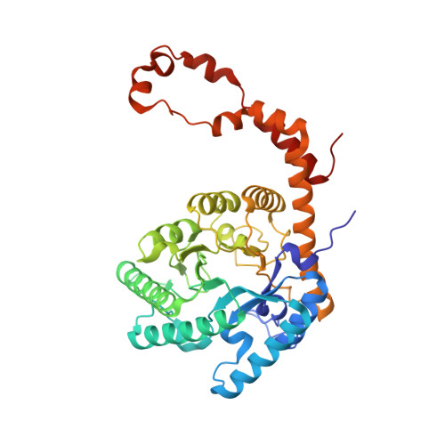
    D-Xylose isomerase (XI) converts the aldo-sugars xylose and glucose to their keto analogs xylulose and fructose, but is strongly inhibited by the polyols xylitol and sorbitol, especially at acidic pH. In order to understand the atomic details of polyol binding to the XI active site, a 2.0 Å resolution room-temperature joint X-ray/neutron structure of XI in complex with Ni(2+) cofactors and sorbitol inhibitor at pH 5.9 and a room-temperature X-ray structure of XI containing Mg(2+) ions and xylitol at the physiological pH of 7.7 were obtained. The protonation of oxygen O5 of the inhibitor, which was found to be deprotonated and negatively charged in previous structures of XI complexed with linear glucose and xylulose, was directly observed. The Ni(2+) ions occupying the catalytic metal site (M2) were found at two locations, while Mg(2+) in M2 is very mobile and has a high B factor. Under acidic conditions sorbitol gains a water-mediated interaction that connects its O1 hydroxyl to Asp257. This contact is not found in structures at basic pH. The new interaction that is formed may improve the binding of the inhibitor, providing an explanation for the increased affinity of the polyols for XI at low pH.


  • Organizational Affiliation

    Bioscience Division, Los Alamos National Laboratory, Los Alamos, NM 87545, USA. ayk@lanl.gov


Macromolecules
Find similar proteins by:  (by identity cutoff)  |  3D Structure
Entity ID: 1
MoleculeChains Sequence LengthOrganismDetailsImage
Xylose isomerase388Streptomyces rubiginosusMutation(s): 0 
EC: 5.3.1.5
UniProt
Find proteins for P24300 (Streptomyces rubiginosus)
Explore P24300 
Go to UniProtKB:  P24300
Entity Groups  
Sequence Clusters30% Identity50% Identity70% Identity90% Identity95% Identity100% Identity
UniProt GroupP24300
Sequence Annotations
Expand
  • Reference Sequence
Experimental Data & Validation

Experimental Data

  • Method: NEUTRON DIFFRACTION
  • Resolution: 2.00 Å
  • R-Value Free: 0.214 
  • R-Value Work: 0.190 
  • R-Value Observed: 0.190 
  • Space Group: I 2 2 2
Unit Cell:
Length ( Å )Angle ( ˚ )
a = 94.061α = 90
b = 99.428β = 90
c = 102.798γ = 90
Software Package:
Software NamePurpose
CNSrefinement
nCNSrefinement
d*TREKdata reduction
d*TREKdata scaling
CNSphasing

Structure Validation

View Full Validation Report


Ligand Structure Quality Assessment 


Entry History 

Deposition Data

Revision History  (Full details and data files)

  • Version 1.0: 2012-08-29
    Type: Initial release
  • Version 1.1: 2012-10-03
    Changes: Database references
  • Version 1.2: 2017-11-15
    Changes: Refinement description
  • Version 1.3: 2020-07-29
    Type: Remediation
    Reason: Carbohydrate remediation
    Changes: Derived calculations, Structure summary
  • Version 1.4: 2024-02-28
    Changes: Data collection, Database references, Structure summary