4DRA

Crystal structure of MHF complex


Experimental Data Snapshot

  • Method: X-RAY DIFFRACTION
  • Resolution: 2.41 Å
  • R-Value Free: 0.242 
  • R-Value Work: 0.190 
  • R-Value Observed: 0.193 

wwPDB Validation   3D Report Full Report


This is version 1.1 of the entry. See complete history


Literature

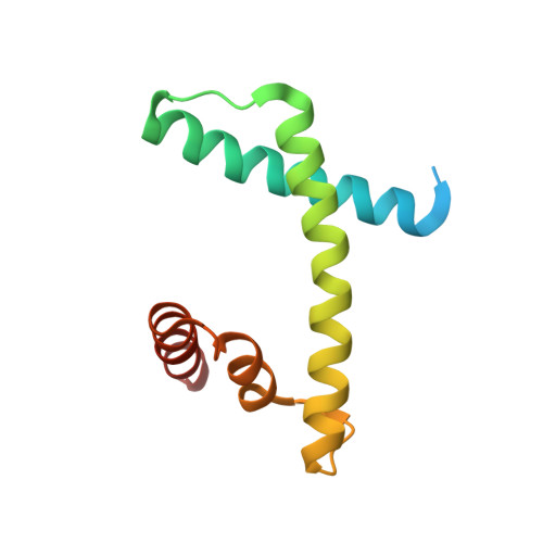
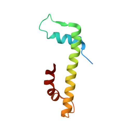
The structure of the FANCM-MHF complex reveals physical features for functional assembly

Tao, Y.Jin, C.Li, X.Qi, S.Chu, L.Niu, L.Yao, X.Teng, M.

(2012) Nat Commun 3: 782-782

  • DOI: https://doi.org/10.1038/ncomms1779
  • Primary Citation of Related Structures:  
    4DRA, 4DRB

  • PubMed Abstract: 

    Fanconi anaemia is a rare genetic disease characterized by chromosomal instability and cancer susceptibility. The Fanconi anaemia complementation group protein M (FANCM) forms an evolutionarily conserved DNA-processing complex with MHF1/MHF2 (histone-fold-containing proteins), which is essential for DNA repair in response to genotoxic stress. Here we present the crystal structures of the MHF1-MHF2 complex alone and bound to a fragment of FANCM (FANCM(661-800), designated FANCM-F). The structures show that MHF1 and MHF2 form a compact tetramer to which FANCM-F binds through a 'dual-V' shaped structure. FANCM-F and (MHF1-MHF2)(2) cooperate to constitute a new DNA-binding site that is coupled to the canonical L1L2 region. Perturbation of the MHF-FANCM-F structural plasticity changes the localization of FANCM in vivo. The MHF-FANCM interaction and its subcellular localization are altered by a disease-associated mutant of FANCM. These findings reveal the molecular basis of MHF-FANCM recognition and provide mechanistic insights into the pathway leading to Fanconi anaemia.


  • Organizational Affiliation

    Key Laboratory of Structural Biology, Chinese Academy of Sciences, Hefei, Anhui 230026, China.


Macromolecules
Find similar proteins by:  (by identity cutoff)  |  3D Structure
Entity ID: 1
MoleculeChains Sequence LengthOrganismDetailsImage
Centromere protein S
A, B, C, D
113Homo sapiensMutation(s): 0 
Gene Names: APITD1CENPSFAAP16MHF1
UniProt & NIH Common Fund Data Resources
Find proteins for Q8N2Z9 (Homo sapiens)
Explore Q8N2Z9 
Go to UniProtKB:  Q8N2Z9
PHAROS:  Q8N2Z9
GTEx:  ENSG00000175279 
Entity Groups  
Sequence Clusters30% Identity50% Identity70% Identity90% Identity95% Identity100% Identity
UniProt GroupQ8N2Z9
Sequence Annotations
Expand
  • Reference Sequence
Find similar proteins by:  (by identity cutoff)  |  3D Structure
Entity ID: 2
MoleculeChains Sequence LengthOrganismDetailsImage
Centromere protein X
E, F, G, H
84Homo sapiensMutation(s): 0 
Gene Names: STRA13CENPXFAAP10MHF2
UniProt & NIH Common Fund Data Resources
Find proteins for A8MT69 (Homo sapiens)
Explore A8MT69 
Go to UniProtKB:  A8MT69
PHAROS:  A8MT69
GTEx:  ENSG00000169689 
Entity Groups  
Sequence Clusters30% Identity50% Identity70% Identity90% Identity95% Identity100% Identity
UniProt GroupA8MT69
Sequence Annotations
Expand
  • Reference Sequence
Experimental Data & Validation

Experimental Data

  • Method: X-RAY DIFFRACTION
  • Resolution: 2.41 Å
  • R-Value Free: 0.242 
  • R-Value Work: 0.190 
  • R-Value Observed: 0.193 
  • Space Group: P 1 21 1
Unit Cell:
Length ( Å )Angle ( ˚ )
a = 41.007α = 90
b = 128.771β = 100.74
c = 88.697γ = 90
Software Package:
Software NamePurpose
HKL-2000data collection
MOLREPphasing
PHENIXrefinement
HKL-2000data reduction
SCALEPACKdata scaling

Structure Validation

View Full Validation Report


Entry History 

Deposition Data

Revision History  (Full details and data files)

  • Version 1.0: 2012-05-16
    Type: Initial release
  • Version 1.1: 2024-03-20
    Changes: Data collection, Database references