Characterisation of the first enzymes committed to lysine biosynthesis in Arabidopsis thaliana
Griffin, M.D.W., Billakanti, J.M., Wason, A., Keller, S., Mertens, H.D.T., Atkinson, S.C., Dobson, R.C.J., Perugini, M.A., Gerrard, J.A., Pearce, F.G.(2012) PLoS One 7: e40318-e40318
- PubMed: 22792278
- DOI: https://doi.org/10.1371/journal.pone.0040318
- Primary Citation of Related Structures:
4DPP, 4DPQ - PubMed Abstract:
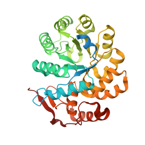
In plants, the lysine biosynthetic pathway is an attractive target for both the development of herbicides and increasing the nutritional value of crops given that lysine is a limiting amino acid in cereals. Dihydrodipicolinate synthase (DHDPS) and dihydrodipicolinate reductase (DHDPR) catalyse the first two committed steps of lysine biosynthesis. Here, we carry out for the first time a comprehensive characterisation of the structure and activity of both DHDPS and DHDPR from Arabidopsis thaliana. The A. thaliana DHDPS enzyme (At-DHDPS2) has similar activity to the bacterial form of the enzyme, but is more strongly allosterically inhibited by (S)-lysine. Structural studies of At-DHDPS2 show (S)-lysine bound at a cleft between two monomers, highlighting the allosteric site; however, unlike previous studies, binding is not accompanied by conformational changes, suggesting that binding may cause changes in protein dynamics rather than large conformation changes. DHDPR from A. thaliana (At-DHDPR2) has similar specificity for both NADH and NADPH during catalysis, and has tighter binding of substrate than has previously been reported. While all known bacterial DHDPR enzymes have a tetrameric structure, analytical ultracentrifugation, and scattering data unequivocally show that At-DHDPR2 exists as a dimer in solution. The exact arrangement of the dimeric protein is as yet unknown, but ab initio modelling of x-ray scattering data is consistent with an elongated structure in solution, which does not correspond to any of the possible dimeric pairings observed in the X-ray crystal structure of DHDPR from other organisms. This increased knowledge of the structure and function of plant lysine biosynthetic enzymes will aid future work aimed at improving primary production.
Organizational Affiliation:
Department of Biochemistry and Molecular Biology, Bio21 Molecular Science and Biotechnology Institute, University of Melbourne, Melbourne, Victoria, Australia.