4DLJ

Human p38 MAP kinase in complex with RL163


Experimental Data Snapshot

  • Method: X-RAY DIFFRACTION
  • Resolution: 2.60 Å
  • R-Value Free: 0.314 
  • R-Value Work: 0.209 
  • R-Value Observed: 0.216 

wwPDB Validation   3D Report Full Report


This is version 1.1 of the entry. See complete history


Literature

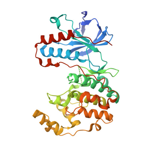
Fluorophore labeled kinase detects ligands that bind within the MAPK insert of p38alpha kinase.

Getlik, M.Simard, J.R.Termathe, M.Grutter, C.Rabiller, M.van Otterlo, W.A.Rauh, D.

(2012) PLoS One 7: e39713-e39713

  • DOI: https://doi.org/10.1371/journal.pone.0039713
  • Primary Citation of Related Structures:  
    4DLI, 4DLJ

  • PubMed Abstract: 

    The vast majority of small molecules known to modulate kinase activity, target the highly conserved ATP-pocket. Consequently, such ligands are often less specific and in case of inhibitors, this leads to the inhibition of multiple kinases. Thus, selective modulation of kinase function remains a major hurdle. One of the next great challenges in kinase research is the identification of ligands which bind to less conserved sites and target the non-catalytic functions of protein kinases. However, approaches that allow for the unambiguous identification of molecules that bind to these less conserved sites are few in number. We have previously reported the use of fluorescent labels in kinases (FLiK) to develop direct kinase binding assays that exclusively detect ligands which stabilize inactive (DFG-out) kinase conformations. Here, we present the successful application of the FLiK approach to develop a high-throughput binding assay capable of directly monitoring ligand binding to a remote site within the MAPK insert of p38α mitogen-activated protein kinase (MAPK). Guided by the crystal structure of an initially identified hit molecule in complex with p38α, we developed a tight binding ligand which may serve as an ideal starting point for further investigations of the biological function of the MAPK insert in regulating the p38α signaling pathway.


  • Organizational Affiliation

    Chemical Genomics Centre of the Max Planck Society, Dortmund, Germany.


Macromolecules
Find similar proteins by:  (by identity cutoff)  |  3D Structure
Entity ID: 1
MoleculeChains Sequence LengthOrganismDetailsImage
Mitogen-activated protein kinase 14360Homo sapiensMutation(s): 0 
Gene Names: MAPK14CSBPCSBP1CSBP2CSPB1MXI2SAPK2A
EC: 2.7.11.24
UniProt & NIH Common Fund Data Resources
Find proteins for Q16539 (Homo sapiens)
Explore Q16539 
Go to UniProtKB:  Q16539
PHAROS:  Q16539
GTEx:  ENSG00000112062 
Entity Groups  
Sequence Clusters30% Identity50% Identity70% Identity90% Identity95% Identity100% Identity
UniProt GroupQ16539
Sequence Annotations
Expand
  • Reference Sequence
Small Molecules
Ligands 1 Unique
IDChains Name / Formula / InChI Key2D Diagram3D Interactions
6RG
Query on 6RG

Download Ideal Coordinates CCD File 
B [auth A],
C [auth A]
2-phenyl-N~4~-(2-phenylethyl)quinazoline-4,7-diamine
C22 H20 N4
TZGHZEALSHYAJC-UHFFFAOYSA-N
Binding Affinity Annotations 
IDSourceBinding Affinity
6RG PDBBind:  4DLJ Kd: 1200 (nM) from 1 assay(s)
Experimental Data & Validation

Experimental Data

  • Method: X-RAY DIFFRACTION
  • Resolution: 2.60 Å
  • R-Value Free: 0.314 
  • R-Value Work: 0.209 
  • R-Value Observed: 0.216 
  • Space Group: P 21 21 21
Unit Cell:
Length ( Å )Angle ( ˚ )
a = 65.98α = 90
b = 74.51β = 90
c = 78.32γ = 90
Software Package:
Software NamePurpose
XSCALEdata scaling
PHASERphasing
REFMACrefinement
PDB_EXTRACTdata extraction
MAR345dtbdata collection

Structure Validation

View Full Validation Report


Entry History 

Deposition Data

Revision History  (Full details and data files)

  • Version 1.0: 2013-01-23
    Type: Initial release
  • Version 1.1: 2023-09-13
    Changes: Data collection, Database references, Derived calculations, Refinement description