Structure and interactions of the cytoplasmic domain of the Yersinia type III secretion protein YscD.
Gamez, A., Mukerjea, R., Alayyoubi, M., Ghassemian, M., Ghosh, P.(2012) J Bacteriol 194: 5949-5958
- PubMed: 22942247
- DOI: https://doi.org/10.1128/JB.00513-12
- Primary Citation of Related Structures:
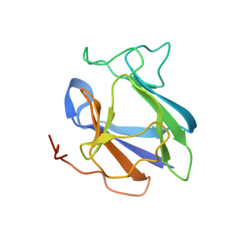
4D9V - PubMed Abstract:
The virulence of a large number of Gram-negative bacterial pathogens depends on the type III secretion (T3S) system, which transports select bacterial proteins into host cells. An essential component of the Yersinia T3S system is YscD, a single-pass inner membrane protein. We report here the 2.52-Å resolution structure of the cytoplasmic domain of YscD, called YscDc. The structure confirms that YscDc consists of a forkhead-associated (FHA) fold, which in many but not all cases specifies binding to phosphothreonine. YscDc, however, lacks the structural properties associated with phosphothreonine binding and thus most likely interacts with partners in a phosphorylation-independent manner. Structural comparison highlighted two loop regions, L3 and L4, as potential sites of interactions. Alanine substitutions at L3 and L4 had no deleterious effects on protein structure or stability but abrogated T3S in a dominant negative manner. To gain insight into the function of L3 and L4, we identified proteins associated with YscD by affinity purification coupled to mass spectrometry. The lipoprotein YscJ was found associated with wild-type YscD, as was the effector YopH. Notably, the L3 and L4 substitution mutants interacted with more YopH than did wild-type YscD. These substitution mutants also interacted with SycH (the specific chaperone for YopH), the putative C-ring component YscQ, and the ruler component YscP, whereas wild-type YscD did not. These results suggest that substitutions in the L3 and L4 loops of YscD disrupted the dissociation of SycH from YopH, leading to the accumulation of a large protein complex that stalled the T3S apparatus.
Organizational Affiliation:
Department of Chemistry & Biochemistry, University of California, San Diego, La Jolla, California, USA.