4CRN

Cryo-EM of a pretermination complex with eRF1 and eRF3


Experimental Data Snapshot

  • Method: ELECTRON MICROSCOPY
  • Resolution: 9.10 Å
  • Aggregation State: PARTICLE 
  • Reconstruction Method: SINGLE PARTICLE 

wwPDB Validation   3D Report Full Report


This is version 1.2 of the entry. See complete history


Literature

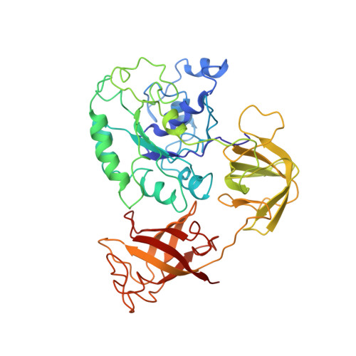
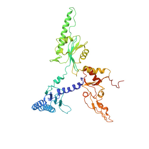
Cryoelectron Microscopic Structures of Eukaryotic Translation Termination Complexes Containing Erf1-Erf3 or Erf1-Abce1.

Preis, A.Heuer, A.Barrio-Garcia, C.Hauser, A.Eyler, D.E.Berninghausen, O.Green, R.Becker, T.Beckmann, R.

(2014) Cell Rep 8: 59

  • DOI: https://doi.org/10.1016/j.celrep.2014.04.058
  • Primary Citation of Related Structures:  
    4CRM, 4CRN

  • PubMed Abstract: 

    Termination and ribosome recycling are essential processes in translation. In eukaryotes, a stop codon in the ribosomal A site is decoded by a ternary complex consisting of release factors eRF1 and guanosine triphosphate (GTP)-bound eRF3. After GTP hydrolysis, eRF3 dissociates, and ABCE1 can bind to eRF1-loaded ribosomes to stimulate peptide release and ribosomal subunit dissociation. Here, we present cryoelectron microscopic (cryo-EM) structures of a pretermination complex containing eRF1-eRF3 and a termination/prerecycling complex containing eRF1-ABCE1. eRF1 undergoes drastic conformational changes: its central domain harboring the catalytically important GGQ loop is either packed against eRF3 or swung toward the peptidyl transferase center when bound to ABCE1. Additionally, in complex with eRF3, the N-terminal domain of eRF1 positions the conserved NIKS motif proximal to the stop codon, supporting its suggested role in decoding, yet it appears to be delocalized in the presence of ABCE1. These results suggest that stop codon decoding and peptide release can be uncoupled during termination.


  • Organizational Affiliation

    Gene Center Munich, Department of Biochemistry, Feodor-Lynen-Strasse 25, University of Munich, 81377 Munich, Germany; Center for Integrated Protein Science Munich, Department of Biochemistry, Feodor-Lynen-Strasse 25, University of Munich, 81377 Munich, Germany.


Macromolecules
Find similar proteins by:  (by identity cutoff)  |  3D Structure
Entity ID: 1
MoleculeChains Sequence LengthOrganismDetailsImage
ERF3 IN RIBOSOME BOUND ERF1-ERF3-GDPNP COMPLEXA [auth P]430Saccharomyces cerevisiaeMutation(s): 0 
UniProt
Find proteins for P05453 (Saccharomyces cerevisiae (strain ATCC 204508 / S288c))
Explore P05453 
Go to UniProtKB:  P05453
Entity Groups  
Sequence Clusters30% Identity50% Identity70% Identity90% Identity95% Identity100% Identity
UniProt GroupP05453
Sequence Annotations
Expand
  • Reference Sequence
Find similar proteins by:  (by identity cutoff)  |  3D Structure
Entity ID: 2
MoleculeChains Sequence LengthOrganismDetailsImage
ERF1 IN RIBOSOME-BOUND ERF1-ERF3-GDPNP COMPLEXB [auth X]437Saccharomyces cerevisiaeMutation(s): 0 
UniProt
Find proteins for P12385 (Saccharomyces cerevisiae (strain ATCC 204508 / S288c))
Explore P12385 
Go to UniProtKB:  P12385
Entity Groups  
Sequence Clusters30% Identity50% Identity70% Identity90% Identity95% Identity100% Identity
UniProt GroupP12385
Sequence Annotations
Expand
  • Reference Sequence
Small Molecules
Ligands 1 Unique
IDChains Name / Formula / InChI Key2D Diagram3D Interactions
GNP
Query on GNP

Download Ideal Coordinates CCD File 
C [auth P]PHOSPHOAMINOPHOSPHONIC ACID-GUANYLATE ESTER
C10 H17 N6 O13 P3
UQABYHGXWYXDTK-UUOKFMHZSA-N
Experimental Data & Validation

Experimental Data

  • Method: ELECTRON MICROSCOPY
  • Resolution: 9.10 Å
  • Aggregation State: PARTICLE 
  • Reconstruction Method: SINGLE PARTICLE 
EM Software:
TaskSoftware PackageVersion
RECONSTRUCTIONSPIDER

Structure Validation

View Full Validation Report


Entry History 

Deposition Data

Revision History  (Full details and data files)

  • Version 1.0: 2014-07-23
    Type: Initial release
  • Version 1.1: 2016-05-11
    Changes: Structure summary
  • Version 1.2: 2017-08-23
    Changes: Data collection