4CLV

Crystal Structure of dodecylphosphocholine-solubilized NccX from Cupriavidus metallidurans 31A


Experimental Data Snapshot

  • Method: X-RAY DIFFRACTION
  • Resolution: 3.12 Å
  • R-Value Free: 0.264 
  • R-Value Work: 0.251 
  • R-Value Observed: 0.252 

wwPDB Validation   3D Report Full Report


Ligand Structure Quality Assessment 


This is version 1.3 of the entry. See complete history


Literature

The X-Ray Structure of Nccx from Cupriavidus Metallidurans 31A Illustrates Potential Dangers of Detergent Solubilization When Generating and Interpreting Crystal Structures of Membrane Proteins.

Ziani, W.Maillard, A.P.Petit-Hartlein, I.Garnier, N.Crouzy, S.Girard, E.Coves, J.

(2014) J Biol Chem 289: 31160

  • DOI: https://doi.org/10.1074/jbc.M114.586537
  • Primary Citation of Related Structures:  
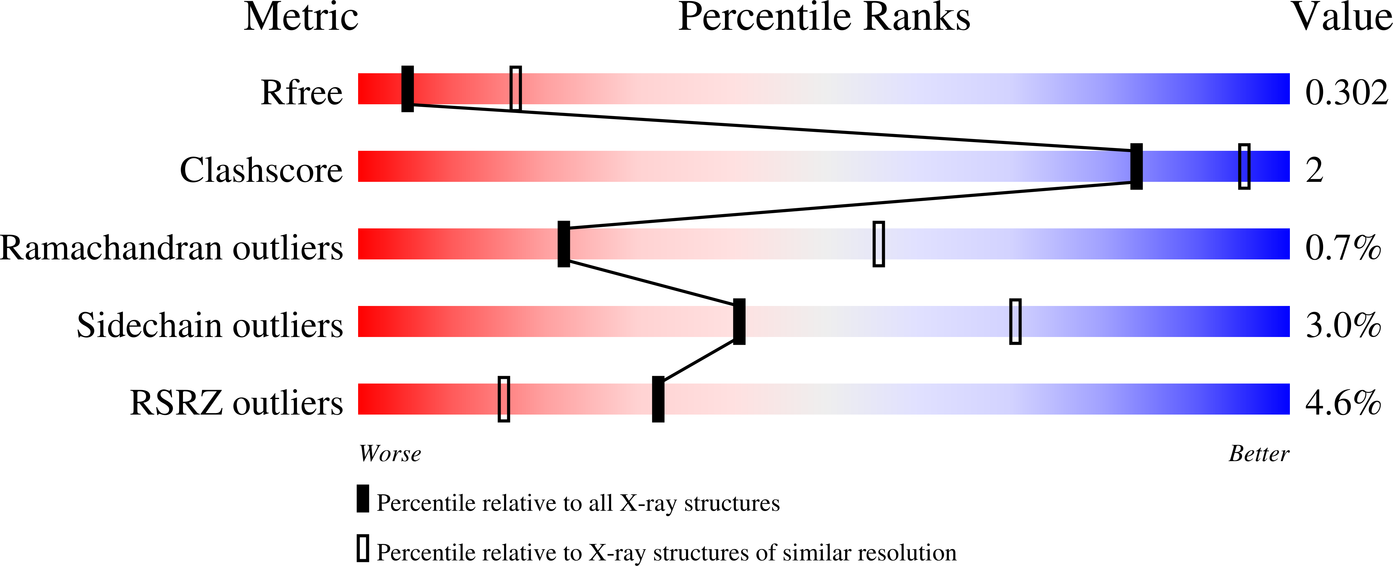
    4CLV

  • PubMed Abstract: 

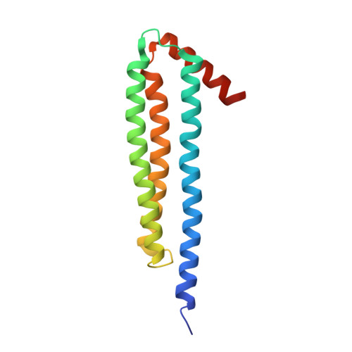
    The x-ray structure of NccX, a type II transmembrane metal sensor, from Cupriavidus metallidurans 31A has been determined at a resolution of 3.12 Å. This was achieved after solubilization by dodecylphosphocholine and purification in the presence of the detergent. NccX crystal structure did not match the model based on the extensively characterized periplasmic domain of its closest homologue CnrX. Instead, the periplasmic domains of NccX appeared collapsed against the hydrophobic transmembrane segments, leading to an aberrant topology incompatible with membrane insertion. This was explained by a detergent-induced redistribution of the hydrophobic interactions among the transmembrane helices and a pair of hydrophobic patches keeping the periplasmic domains together in the native dimer. Molecular dynamics simulations performed with the full-length protein or with the transmembrane segments were used along with in vivo homodimerization assays (TOXCAT) to evaluate the determinants of the interactions between NccX protomers. Taken as a whole, computational and experimental results are in agreement with the structural model of CnrX where a cradle-shaped periplasmic metal sensor domain is anchored into the inner membrane by two N-terminal helices. In addition, they show that the main determinant of NccX dimerization is the periplasmic soluble domain and that the interaction between transmembrane segments is highly dynamic. The present work introduces a new crystal structure for a transmembrane protein and, in line with previous studies, substantiates the use of complementary theoretical and in vivo investigations to rationalize a three-dimensional structure obtained in non-native conditions.


  • Organizational Affiliation

    From the Institut de Biologie Structurale, UMR 5075 CNRS-Commissariat à l'Energie Atomique (CEA)-Université Grenoble-Alpes, 71 Avenue des Martyrs, 38044 Grenoble Cedex 9, France.


Macromolecules
Find similar proteins by:  (by identity cutoff)  |  3D Structure
Entity ID: 1
MoleculeChains Sequence LengthOrganismDetailsImage
NICKEL-COBALT-CADMIUM RESISTANCE PROTEIN NCCX
A, B
148Cupriavidus metalliduransMutation(s): 0 
UniProt
Find proteins for Q44582 (Alcaligenes xylosoxydans xylosoxydans)
Explore Q44582 
Go to UniProtKB:  Q44582
Entity Groups  
Sequence Clusters30% Identity50% Identity70% Identity90% Identity95% Identity100% Identity
UniProt GroupQ44582
Sequence Annotations
Expand
  • Reference Sequence
Small Molecules
Ligands 3 Unique
IDChains Name / Formula / InChI Key2D Diagram3D Interactions
PC
Query on PC

Download Ideal Coordinates CCD File 
K [auth A],
R [auth B]
PHOSPHOCHOLINE
C5 H15 N O4 P
YHHSONZFOIEMCP-UHFFFAOYSA-O
PO4
Query on PO4

Download Ideal Coordinates CCD File 
I [auth A],
J [auth A],
P [auth B],
Q [auth B]
PHOSPHATE ION
O4 P
NBIIXXVUZAFLBC-UHFFFAOYSA-K
ZN
Query on ZN

Download Ideal Coordinates CCD File 
C [auth A]
D [auth A]
E [auth A]
F [auth A]
G [auth A]
C [auth A],
D [auth A],
E [auth A],
F [auth A],
G [auth A],
H [auth A],
L [auth B],
M [auth B],
N [auth B],
O [auth B]
ZINC ION
Zn
PTFCDOFLOPIGGS-UHFFFAOYSA-N
Experimental Data & Validation

Experimental Data

  • Method: X-RAY DIFFRACTION
  • Resolution: 3.12 Å
  • R-Value Free: 0.264 
  • R-Value Work: 0.251 
  • R-Value Observed: 0.252 
  • Space Group: P 43 21 2
Unit Cell:
Length ( Å )Angle ( ˚ )
a = 53.8α = 90
b = 53.8β = 90
c = 300.04γ = 90
Software Package:
Software NamePurpose
BUSTERrefinement
XDSdata reduction
XSCALEdata scaling
PHASERphasing

Structure Validation

View Full Validation Report


Ligand Structure Quality Assessment 


Entry History 

Deposition Data

Revision History  (Full details and data files)

  • Version 1.0: 2014-10-01
    Type: Initial release
  • Version 1.1: 2014-10-08
    Changes: Database references
  • Version 1.2: 2014-11-19
    Changes: Database references
  • Version 1.3: 2015-03-25
    Changes: Atomic model