4CFE

Structure of full length human AMPK in complex with a small molecule activator, a benzimidazole derivative (991)


Experimental Data Snapshot

  • Method: X-RAY DIFFRACTION
  • Resolution: 3.02 Å
  • R-Value Free: 0.253 
  • R-Value Work: 0.218 
  • R-Value Observed: 0.219 

wwPDB Validation   3D Report Full Report


Ligand Structure Quality Assessment 


This is version 1.4 of the entry. See complete history


Literature

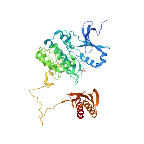
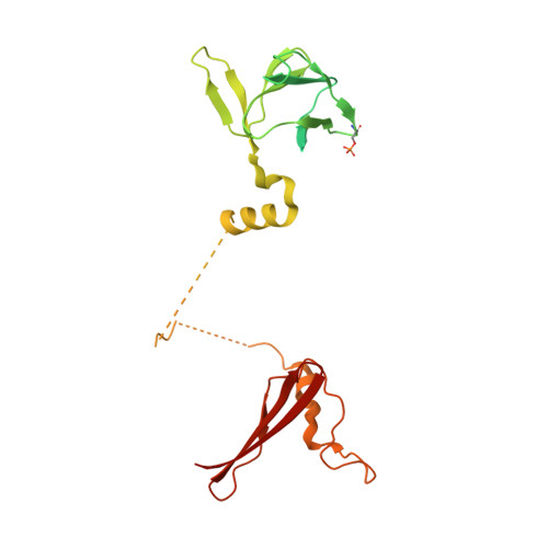
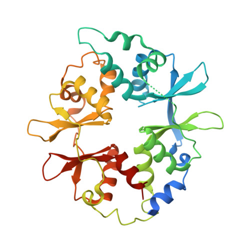
Structural Basis of Ampk Regulation by Small Molecule Activators.

Xiao, B.Sanders, M.J.Carmena, D.Bright, N.J.Haire, L.F.Underwood, E.Patel, B.R.Heath, R.B.Walker, P.A.Hallen, S.Giordanetto, F.Martin, S.R.Carling, D.Gamblin, S.J.

(2013) Nat Commun 4: 3017

  • DOI: https://doi.org/10.1038/ncomms4017
  • Primary Citation of Related Structures:  
    4CFE, 4CFF

  • PubMed Abstract: 

    AMP-activated protein kinase (AMPK) plays a major role in regulating cellular energy balance by sensing and responding to increases in AMP/ADP concentration relative to ATP. Binding of AMP causes allosteric activation of the enzyme and binding of either AMP or ADP promotes and maintains the phosphorylation of threonine 172 within the activation loop of the kinase. AMPK has attracted widespread interest as a potential therapeutic target for metabolic diseases including type 2 diabetes and, more recently, cancer. A number of direct AMPK activators have been reported as having beneficial effects in treating metabolic diseases, but there has been no structural basis for activator binding to AMPK. Here we present the crystal structure of human AMPK in complex with a small molecule activator that binds at a site between the kinase domain and the carbohydrate-binding module, stabilising the interaction between these two components. The nature of the activator-binding pocket suggests the involvement of an additional, as yet unidentified, metabolite in the physiological regulation of AMPK. Importantly, the structure offers new opportunities for the design of small molecule activators of AMPK for treatment of metabolic disorders.


  • Organizational Affiliation

    1] MRC National Institute for Medical Research, The Ridgeway, Mill Hill, London NW7 1AA, UK [2].


Macromolecules
Find similar proteins by:  (by identity cutoff)  |  3D Structure
Entity ID: 1
MoleculeChains Sequence LengthOrganismDetailsImage
5'-AMP-ACTIVATED PROTEIN KINASE CATALYTIC SUBUNIT ALPHA-2
A, C
571Homo sapiensMutation(s): 0 
EC: 2.7.11.1 (PDB Primary Data), 2.7.11.27 (PDB Primary Data), 2.7.11.31 (PDB Primary Data)
UniProt & NIH Common Fund Data Resources
Find proteins for P54646 (Homo sapiens)
Explore P54646 
Go to UniProtKB:  P54646
PHAROS:  P54646
GTEx:  ENSG00000162409 
Entity Groups  
Sequence Clusters30% Identity50% Identity70% Identity90% Identity95% Identity100% Identity
UniProt GroupP54646
Sequence Annotations
Expand
  • Reference Sequence
Find similar proteins by:  (by identity cutoff)  |  3D Structure
Entity ID: 2
MoleculeChains Sequence LengthOrganismDetailsImage
5'-AMP-ACTIVATED PROTEIN KINASE SUBUNIT BETA-1
B, D
286Homo sapiensMutation(s): 0 
UniProt & NIH Common Fund Data Resources
Find proteins for Q9Y478 (Homo sapiens)
Explore Q9Y478 
Go to UniProtKB:  Q9Y478
PHAROS:  Q9Y478
GTEx:  ENSG00000111725 
Entity Groups  
Sequence Clusters30% Identity50% Identity70% Identity90% Identity95% Identity100% Identity
UniProt GroupQ9Y478
Sequence Annotations
Expand
  • Reference Sequence
Find similar proteins by:  (by identity cutoff)  |  3D Structure
Entity ID: 3
MoleculeChains Sequence LengthOrganismDetailsImage
5'-AMP-ACTIVATED PROTEIN KINASE SUBUNIT GAMMA-1
E, F
331Homo sapiensMutation(s): 0 
UniProt & NIH Common Fund Data Resources
Find proteins for P54619 (Homo sapiens)
Explore P54619 
Go to UniProtKB:  P54619
PHAROS:  P54619
GTEx:  ENSG00000181929 
Entity Groups  
Sequence Clusters30% Identity50% Identity70% Identity90% Identity95% Identity100% Identity
UniProt GroupP54619
Sequence Annotations
Expand
  • Reference Sequence
Small Molecules
Ligands 3 Unique
IDChains Name / Formula / InChI Key2D Diagram3D Interactions
STU
Query on STU

Download Ideal Coordinates CCD File 
G [auth A],
I [auth C]
STAUROSPORINE
C28 H26 N4 O3
HKSZLNNOFSGOKW-FYTWVXJKSA-N
992
Query on 992

Download Ideal Coordinates CCD File 
H [auth A],
J [auth C]
5-[[6-chloranyl-5-(1-methylindol-5-yl)-1H-benzimidazol-2-yl]oxy]-2-methyl-benzoic acid
C24 H18 Cl N3 O3
FHWSAZXFPUMKFL-UHFFFAOYSA-N
AMP
Query on AMP

Download Ideal Coordinates CCD File 
K [auth E]
L [auth E]
M [auth E]
N [auth F]
O [auth F]
K [auth E],
L [auth E],
M [auth E],
N [auth F],
O [auth F],
P [auth F]
ADENOSINE MONOPHOSPHATE
C10 H14 N5 O7 P
UDMBCSSLTHHNCD-KQYNXXCUSA-N
Modified Residues  2 Unique
IDChains TypeFormula2D DiagramParent
TPO
Query on TPO
A, C
L-PEPTIDE LINKINGC4 H10 N O6 PTHR
SEP
Query on SEP
B, D
L-PEPTIDE LINKINGC3 H8 N O6 PSER
Binding Affinity Annotations 
IDSourceBinding Affinity
992 PDBBind:  4CFE Kd: 60 (nM) from 1 assay(s)
BindingDB:  4CFE EC50: 3 (nM) from 1 assay(s)
Experimental Data & Validation

Experimental Data

  • Method: X-RAY DIFFRACTION
  • Resolution: 3.02 Å
  • R-Value Free: 0.253 
  • R-Value Work: 0.218 
  • R-Value Observed: 0.219 
  • Space Group: P 1 21 1
Unit Cell:
Length ( Å )Angle ( ˚ )
a = 76.03α = 90
b = 134.141β = 92.42
c = 140.558γ = 90
Software Package:
Software NamePurpose
PHENIXrefinement
DENZOdata reduction
SCALEPACKdata scaling
PHASERphasing

Structure Validation

View Full Validation Report


Ligand Structure Quality Assessment 


Entry History 

Revision History  (Full details and data files)

  • Version 1.0: 2013-12-25
    Type: Initial release
  • Version 1.1: 2014-01-08
    Changes: Database references
  • Version 1.2: 2014-02-12
    Changes: Data collection, Refinement description
  • Version 1.3: 2019-05-08
    Changes: Advisory, Data collection, Derived calculations, Experimental preparation, Other
  • Version 1.4: 2023-12-20
    Changes: Advisory, Data collection, Database references, Derived calculations, Other, Refinement description