4CEG

Crystal structure of Aurora A 122-403 C290A, C393A bound to ADP


Experimental Data Snapshot

  • Method: X-RAY DIFFRACTION
  • Resolution: 2.10 Å
  • R-Value Free: 0.237 
  • R-Value Work: 0.200 
  • R-Value Observed: 0.202 

wwPDB Validation   3D Report Full Report


Ligand Structure Quality Assessment 


This is version 1.5 of the entry. See complete history


Literature

The Structure of C290A:C393A Aurora a Provides Structural Insights Into Kinase Regulation.

Burgess, S.G.Bayliss, R.

(2015) Acta Crystallogr Sect F Struct Biol Cryst Commun 71: 315

  • DOI: https://doi.org/10.1107/S2053230X15002290
  • Primary Citation of Related Structures:  
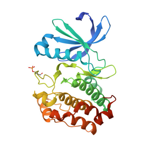
    4CEG

  • PubMed Abstract: 

    Aurora A is a Ser/Thr protein kinase that functions in cell-cycle regulation and is implicated in cancer development. During mitosis, Aurora A is activated by autophosphorylation on its activation loop at Thr288. The Aurora A catalytic domain (amino acids 122-403) expressed in Escherichia coli autophosphorylates on two activation-loop threonine residues (Thr288 and Thr287), whereas a C290A,C393A double point mutant of the Aurora A catalytic domain autophosphorylates only on Thr288. The structure of the complex of this mutant with ADP and magnesium was determined to 2.1 Å resolution using molecular replacement. This is an improvement on the existing 2.75 Å resolution structure of the equivalent wild-type complex. The structure confirms that single phosphorylation of the activation loop on Thr288 is insufficient to stabilize a `fully active' conformation of the activation loop in the absence of binding to TPX2.


  • Organizational Affiliation

    Department of Biochemistry, University of Leicester, Lancaster Road, Leicester LE1 9HN, England.


Macromolecules
Find similar proteins by:  (by identity cutoff)  |  3D Structure
Entity ID: 1
MoleculeChains Sequence LengthOrganismDetailsImage
AURORA KINASE A285Homo sapiensMutation(s): 2 
UniProt & NIH Common Fund Data Resources
Find proteins for O14965 (Homo sapiens)
Explore O14965 
Go to UniProtKB:  O14965
PHAROS:  O14965
GTEx:  ENSG00000087586 
Entity Groups  
Sequence Clusters30% Identity50% Identity70% Identity90% Identity95% Identity100% Identity
UniProt GroupO14965
Sequence Annotations
Expand
  • Reference Sequence
Small Molecules
Ligands 4 Unique
IDChains Name / Formula / InChI Key2D Diagram3D Interactions
ADP
Query on ADP

Download Ideal Coordinates CCD File 
B [auth A]ADENOSINE-5'-DIPHOSPHATE
C10 H15 N5 O10 P2
XTWYTFMLZFPYCI-KQYNXXCUSA-N
GOL
Query on GOL

Download Ideal Coordinates CCD File 
C [auth A],
D [auth A]
GLYCEROL
C3 H8 O3
PEDCQBHIVMGVHV-UHFFFAOYSA-N
CL
Query on CL

Download Ideal Coordinates CCD File 
G [auth A],
H [auth A],
I [auth A],
J [auth A]
CHLORIDE ION
Cl
VEXZGXHMUGYJMC-UHFFFAOYSA-M
MG
Query on MG

Download Ideal Coordinates CCD File 
E [auth A],
F [auth A]
MAGNESIUM ION
Mg
JLVVSXFLKOJNIY-UHFFFAOYSA-N
Modified Residues  1 Unique
IDChains TypeFormula2D DiagramParent
TPO
Query on TPO
A
L-PEPTIDE LINKINGC4 H10 N O6 PTHR
Experimental Data & Validation

Experimental Data

  • Method: X-RAY DIFFRACTION
  • Resolution: 2.10 Å
  • R-Value Free: 0.237 
  • R-Value Work: 0.200 
  • R-Value Observed: 0.202 
  • Space Group: P 61 2 2
Unit Cell:
Length ( Å )Angle ( ˚ )
a = 80.95α = 90
b = 80.95β = 90
c = 174.53γ = 120
Software Package:
Software NamePurpose
PHENIXrefinement
xia2data reduction
xia2data scaling
PHENIXphasing

Structure Validation

View Full Validation Report


Ligand Structure Quality Assessment 


Entry History 

Deposition Data

Revision History  (Full details and data files)

  • Version 1.0: 2015-01-28
    Type: Initial release
  • Version 1.1: 2015-03-04
    Changes: Database references
  • Version 1.2: 2015-03-11
    Changes: Database references
  • Version 1.3: 2015-03-25
    Changes: Database references
  • Version 1.4: 2019-03-06
    Changes: Data collection, Derived calculations, Experimental preparation, Other
  • Version 1.5: 2023-12-20
    Changes: Data collection, Database references, Derived calculations, Other, Refinement description