4C1L

Crystal structure of pyrococcus furiosus 3-deoxy-D-arabino- heptulosonate 7-phosphate synthase I181D interface mutant


Experimental Data Snapshot

  • Method: X-RAY DIFFRACTION
  • Resolution: 1.80 Å
  • R-Value Free: 0.183 
  • R-Value Work: 0.164 
  • R-Value Observed: 0.165 

wwPDB Validation   3D Report Full Report


This is version 1.4 of the entry. See complete history


Literature

Destabilization of the homotetrameric assembly of 3-deoxy-D-arabino-heptulosonate-7-phosphate synthase from the hyperthermophile Pyrococcus furiosus enhances enzymatic activity.

Nazmi, A.R.Schofield, L.R.Dobson, R.C.Jameson, G.B.Parker, E.J.

(2014) J Mol Biol 426: 656-673

  • DOI: https://doi.org/10.1016/j.jmb.2013.11.008
  • Primary Citation of Related Structures:  
    4C1K, 4C1L

  • PubMed Abstract: 

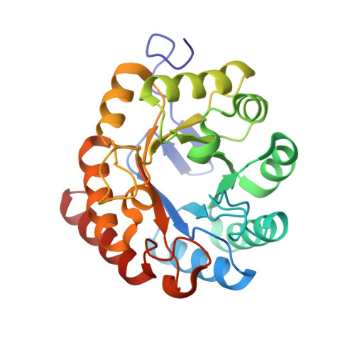
    Many proteins adopt homomeric quaternary structures to support their biological function, including the first enzyme of the shikimate pathway that is ultimately responsible for the biosynthesis of the aromatic amino acids in plants and microorganisms. This enzyme, 3-deoxy-d-arabino-heptulosonate-7-phosphate synthase (DAH7PS), adopts a variety of different quaternary structures depending on the organism in which it is found. The DAH7PS from the hyperthermophilic archaebacterium Pyrococcus furiosus was previously shown to be tetrameric in its crystalline form, and this quaternary association is confirmed in an improved structure in a different crystal system. This tetramer is also present in solution as revealed by small-angle X-ray scattering and analytical ultracentrifugation. This homotetrameric form has two distinct interfaces, both of which bury over 10% each of the surface area of a single monomer. Substitution of Ile for Asp in the hydrophobic region of one interface gives a protein with a remarkable 4-fold higher maximum catalytic rate than the wild-type enzyme. Analytical ultracentrifugation at pH7.5 reveals that the tetrameric form is destabilized; although the protein crystallizes as a tetramer, equilibrium exists between tetrameric and dimeric forms with a dissociation constant of 22 μM. Thus, under the conditions of kinetic assay, the enzyme is primarily dimeric, revealing that the dimeric form is a fully functional catalyst. However, in comparison to the wild-type protein, the thermal stability of the dimeric protein is significantly compromised. Thus, an unusual compromise of enzymatic activity versus stability is observed for this DAH7PS from an organism that favors a hyperthermophilic environment.


  • Organizational Affiliation

    Biomolecular Interaction Centre and Department of Chemistry, University of Canterbury, PO Box 4800, Christchurch 8140, New Zealand.


Macromolecules
Find similar proteins by:  (by identity cutoff)  |  3D Structure
Entity ID: 1
MoleculeChains Sequence LengthOrganismDetailsImage
2-dehydro-3-deoxyphosphoheptonate aldolase
A, B
262Pyrococcus furiosusMutation(s): 1 
Gene Names: PF1690
EC: 2.5.1.54
UniProt
Find proteins for Q8U0A9 (Pyrococcus furiosus (strain ATCC 43587 / DSM 3638 / JCM 8422 / Vc1))
Explore Q8U0A9 
Go to UniProtKB:  Q8U0A9
Entity Groups  
Sequence Clusters30% Identity50% Identity70% Identity90% Identity95% Identity100% Identity
UniProt GroupQ8U0A9
Sequence Annotations
Expand
  • Reference Sequence
Small Molecules
Ligands 4 Unique
IDChains Name / Formula / InChI Key2D Diagram3D Interactions
1PE
Query on 1PE

Download Ideal Coordinates CCD File 
G [auth B]PENTAETHYLENE GLYCOL
C10 H22 O6
JLFNLZLINWHATN-UHFFFAOYSA-N
MN
Query on MN

Download Ideal Coordinates CCD File 
C [auth A],
F [auth B]
MANGANESE (II) ION
Mn
WAEMQWOKJMHJLA-UHFFFAOYSA-N
K
Query on K

Download Ideal Coordinates CCD File 
E [auth A],
K [auth B]
POTASSIUM ION
K
NPYPAHLBTDXSSS-UHFFFAOYSA-N
CL
Query on CL

Download Ideal Coordinates CCD File 
D [auth A],
H [auth B],
I [auth B],
J [auth B]
CHLORIDE ION
Cl
VEXZGXHMUGYJMC-UHFFFAOYSA-M
Experimental Data & Validation

Experimental Data

  • Method: X-RAY DIFFRACTION
  • Resolution: 1.80 Å
  • R-Value Free: 0.183 
  • R-Value Work: 0.164 
  • R-Value Observed: 0.165 
  • Space Group: I 2 2 2
Unit Cell:
Length ( Å )Angle ( ˚ )
a = 87.942α = 90
b = 110.923β = 90
c = 143.531γ = 90
Software Package:
Software NamePurpose
REFMACrefinement
XDSdata reduction
SCALAdata scaling
PHASERphasing

Structure Validation

View Full Validation Report


Entry History 

Deposition Data

Revision History  (Full details and data files)

  • Version 1.0: 2013-11-20
    Type: Initial release
  • Version 1.1: 2013-11-27
    Changes: Database references
  • Version 1.2: 2014-02-05
    Changes: Database references
  • Version 1.3: 2018-09-19
    Changes: Data collection, Database references, Source and taxonomy, Structure summary
  • Version 1.4: 2023-12-20
    Changes: Data collection, Database references, Derived calculations, Other, Refinement description