


Exploring the Surface Determinants of Paracoccus Pantotrophus Pseudoazurin
Freire, F., Mestre, A., Pinho, J., Najmudin, S., Bonifacio, C., Pauleta, S.R., Romao, M.J.To be published.
Experimental Data Snapshot
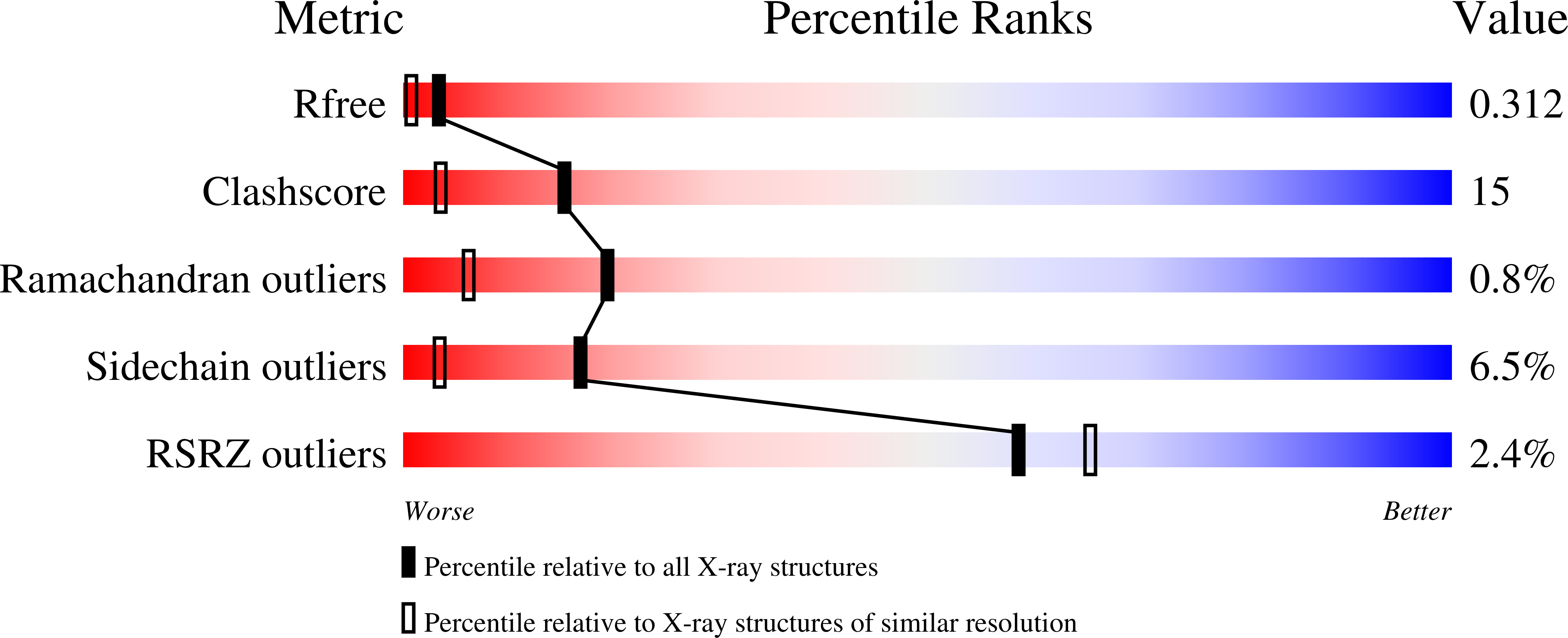
wwPDB Validation 3D Report Full Report
Entity ID: 1 | |||||
|---|---|---|---|---|---|
| Molecule | Chains | Sequence Length | Organism | Details | Image |
| PSEUDOAZURIN | 123 | Paracoccus pantotrophus | Mutation(s): 1 | 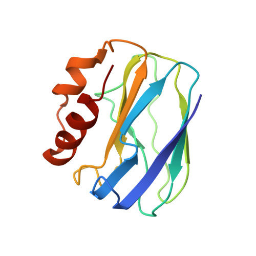 | |
UniProt | |||||
Find proteins for P80401 (Paracoccus pantotrophus) Explore P80401 Go to UniProtKB: P80401 | |||||
Entity Groups | |||||
| Sequence Clusters | 30% Identity50% Identity70% Identity90% Identity95% Identity100% Identity | ||||
| UniProt Group | P80401 | ||||
Sequence AnnotationsExpand | |||||
| |||||
| Ligands 2 Unique | |||||
|---|---|---|---|---|---|
| ID | Chains | Name / Formula / InChI Key | 2D Diagram | 3D Interactions | |
| SO4 Query on SO4 | D [auth A], E [auth A], G [auth B], H [auth B] | SULFATE ION O4 S QAOWNCQODCNURD-UHFFFAOYSA-L |  | ||
| CU Query on CU | C [auth A], F [auth B] | COPPER (II) ION Cu JPVYNHNXODAKFH-UHFFFAOYSA-N |  | ||
| Length ( Å ) | Angle ( ˚ ) |
|---|---|
| a = 41.31 | α = 90 |
| b = 71.72 | β = 90 |
| c = 91.7 | γ = 90 |
| Software Name | Purpose |
|---|---|
| PHENIX | refinement |
| iMOSFLM | data reduction |
| Aimless | data scaling |
| PHASER | phasing |
RCSB PDB (citation) is hosted by
RCSB PDB is a member of the

