4BQ5

Structural analysis of an exo-beta-agarase


Experimental Data Snapshot

  • Method: X-RAY DIFFRACTION
  • Resolution: 2.30 Å
  • R-Value Free: 0.251 
  • R-Value Work: 0.189 
  • R-Value Observed: 0.192 

wwPDB Validation   3D Report Full Report


This is version 2.1 of the entry. See complete history


Literature

Substrate Recognition and Hydrolysis by a Family 50 Exo-Beta-Agarase Aga50D from the Marine Bacterium Saccharophagus Degradans

Pluvinage, B.Hehemann, J.H.Boraston, A.B.

(2013) J Biol Chem 288: 28078

  • DOI: https://doi.org/10.1074/jbc.M113.491068
  • Primary Citation of Related Structures:  
    4BQ2, 4BQ3, 4BQ4, 4BQ5

  • PubMed Abstract: 

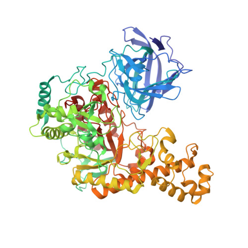
    The bacteria that metabolize agarose use multiple enzymes of complementary specificities to hydrolyze the glycosidic linkages in agarose, a linear polymer comprising the repeating disaccharide subunit of neoagarobiose (3,6-anhydro-l-galactose-α-(1,3)-d-galactose) that are β-(1,4)-linked. Here we present the crystal structure of a glycoside hydrolase family 50 exo-β-agarase, Aga50D, from the marine microbe Saccharophagus degradans. This enzyme catalyzes a critical step in the metabolism of agarose by S. degradans through cleaving agarose oligomers into neoagarobiose products that can be further processed into monomers. The crystal structure of Aga50D to 1.9 Å resolution reveals a (β/α)8-barrel fold that is elaborated with a β-sandwich domain and extensive loops. The structures of catalytically inactivated Aga50D in complex with non-hydrolyzed neoagarotetraose (2.05 Å resolution) and neoagarooctaose (2.30 Å resolution) provide views of Michaelis complexes for a β-agarase. In these structures, the d-galactose residue in the -1 subsite is distorted into a (1)S3 skew boat conformation. The relative positioning of the putative catalytic residues are most consistent with a retaining catalytic mechanism. Additionally, the neoagarooctaose complex showed that this extended substrate made substantial interactions with the β-sandwich domain, which resembles a carbohydrate-binding module, thus creating additional plus (+) subsites and funneling the polymeric substrate through the tunnel-shaped active site. A synthesis of these results in combination with an additional neoagarobiose product complex suggests a potential exo-processive mode of action of Aga50D on the agarose double helix.


  • Organizational Affiliation

    From the Department of Biochemistry and Microbiology, University of Victoria, Victoria, British Columbia V8W 3P6, Canada and.


Macromolecules
Find similar proteins by:  (by identity cutoff)  |  3D Structure
Entity ID: 1
MoleculeChains Sequence LengthOrganismDetailsImage
B-AGARASE
A, B
750Saccharophagus degradans 2-40Mutation(s): 1 
EC: 3.2.1.81
UniProt
Find proteins for Q21HC5 (Saccharophagus degradans (strain 2-40 / ATCC 43961 / DSM 17024))
Explore Q21HC5 
Go to UniProtKB:  Q21HC5
Entity Groups  
Sequence Clusters30% Identity50% Identity70% Identity90% Identity95% Identity100% Identity
UniProt GroupQ21HC5
Sequence Annotations
Expand
  • Reference Sequence
Oligosaccharides

Help

Entity ID: 2
MoleculeChains Length2D Diagram Glycosylation3D Interactions
3,6-anhydro-alpha-L-galactopyranose-(1-3)-beta-D-galactopyranose
C, D, E, G, H
2N/A
Glycosylation Resources
GlyTouCan:  G45207BN
GlyCosmos:  G45207BN
Entity ID: 3
MoleculeChains Length2D Diagram Glycosylation3D Interactions
3,6-anhydro-alpha-L-galactopyranose-(1-3)-beta-D-galactopyranose-(1-4)-3,6-anhydro-alpha-L-galactopyranose-(1-3)-beta-D-galactopyranose
F
4N/A
Glycosylation Resources
GlyTouCan:  G46404PO
GlyCosmos:  G46404PO
Small Molecules
Ligands 4 Unique
IDChains Name / Formula / InChI Key2D Diagram3D Interactions
GAL
Query on GAL

Download Ideal Coordinates CCD File 
J [auth A]beta-D-galactopyranose
C6 H12 O6
WQZGKKKJIJFFOK-FPRJBGLDSA-N
AAL
Query on AAL

Download Ideal Coordinates CCD File 
I [auth A]3,6-anhydro-alpha-L-galactopyranose
C6 H10 O5
DCQFFOLNJVGHLW-DSOBHZJASA-N
GOL
Query on GOL

Download Ideal Coordinates CCD File 
K [auth A],
O [auth B],
P [auth B],
Q [auth B]
GLYCEROL
C3 H8 O3
PEDCQBHIVMGVHV-UHFFFAOYSA-N
CA
Query on CA

Download Ideal Coordinates CCD File 
L [auth A]
M [auth A]
N [auth A]
R [auth B]
S [auth B]
L [auth A],
M [auth A],
N [auth A],
R [auth B],
S [auth B],
T [auth B]
CALCIUM ION
Ca
BHPQYMZQTOCNFJ-UHFFFAOYSA-N
Experimental Data & Validation

Experimental Data

  • Method: X-RAY DIFFRACTION
  • Resolution: 2.30 Å
  • R-Value Free: 0.251 
  • R-Value Work: 0.189 
  • R-Value Observed: 0.192 
  • Space Group: P 21 21 21
Unit Cell:
Length ( Å )Angle ( ˚ )
a = 69.03α = 90
b = 116.17β = 90
c = 207.97γ = 90
Software Package:
Software NamePurpose
REFMACrefinement
MOSFLMdata reduction
SCALAdata scaling
PHASERphasing

Structure Validation

View Full Validation Report


Entry History 

Deposition Data

Revision History  (Full details and data files)

  • Version 1.0: 2013-08-14
    Type: Initial release
  • Version 1.1: 2013-08-21
    Changes: Database references
  • Version 1.2: 2013-08-28
    Changes: Atomic model, Data collection, Derived calculations
  • Version 1.3: 2013-10-16
    Changes: Database references
  • Version 1.4: 2020-07-01
    Changes: Advisory, Data collection, Derived calculations, Other
  • Version 2.0: 2020-07-29
    Type: Remediation
    Reason: Carbohydrate remediation
    Changes: Advisory, Atomic model, Data collection, Derived calculations, Structure summary
  • Version 2.1: 2023-12-20
    Changes: Advisory, Data collection, Database references, Refinement description, Structure summary