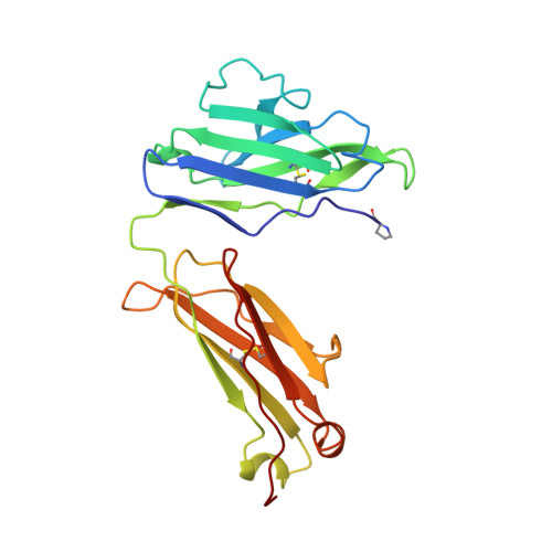
Three quaternary structures for a single protein.
Huang, D.B., Ainsworth, C.F., Stevens, F.J., Schiffer, M.(1996) Proc Natl Acad Sci U S A 93: 7017-7021
- PubMed: 8692936
- DOI: https://doi.org/10.1073/pnas.93.14.7017
- Primary Citation of Related Structures:
1BJM, 3BJL, 4BJL - PubMed Abstract:
The structure of a multisubunit protein (immunoglobulin light chain) was solved in three crystal forms, differing only in the solvent of crystallization. The three structures were obtained at high ionic strength and low pH, high ionic strength and high pH, and low ionic strength and neutral pH. The three resulting "snapshots" of possible structures show that their variable-domain interactions differ, reflecting their stabilities under specific solvent conditions. In the three crystal forms, the variable domains had different rotational and translational relationships, whereas no alteration of the constant domains was found. The critical residues involved in the observed effect of the solvent are tryptophans and histidines located between the two variable domains in the dimeric structure. Tryptophan residues are commonly found in interfaces between proteins and their subunits, and histidines have been implicated in pH-dependent conformation changes. The quaternary structure observed for a multisubunit protein or protein complex in a crystal may be influenced by the interactions of the constituents within the molecule or complex and/or by crystal packing interactions. The comparison of buried surface areas and hydrogen bonds between the domains forming the molecule and between the molecules forming the crystals suggest that, for this system, the interactions within the molecule are most likely the determining factors.
Organizational Affiliation:
Center for Mechanistic Biology and Biotechnology, Argonne National Laboratory, Argonne, IL 60439-4833, USA.