4BEG

Structure of Rv2140c, a phosphatidyl-ethanolamine binding protein from Mycobacterium tuberculosis in complex with sulphate


Experimental Data Snapshot

  • Method: X-RAY DIFFRACTION
  • Resolution: 1.42 Å
  • R-Value Free: 0.195 
  • R-Value Work: 0.149 
  • R-Value Observed: 0.151 

wwPDB Validation   3D Report Full Report


This is version 1.4 of the entry. See complete history


Literature

Structural and Biochemical Characterization of Rv2140C, a Phosphatidylethanolamine-Binding Protein from Mycobacterium Tuberculosis.

Eulenburg, G.Higman, V.A.Diehl, A.Wilmanns, M.Holton, S.J.

(2013) FEBS Lett 587: 2936

  • DOI: https://doi.org/10.1016/j.febslet.2013.07.038
  • Primary Citation of Related Structures:  
    4BEG

  • PubMed Abstract: 

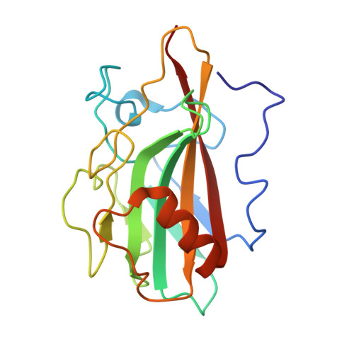
    Rv2140c is one of many conserved Mycobacterium tuberculosis proteins for which no molecular function has been identified. We have determined a high-resolution crystal structure of the Rv2140c gene product, which reveals a dimeric complex that shares strong structural homology with the phosphatidylethanolamine-binding family of proteins. Rv2140c forms low-millimolar interactions with a selection of soluble phosphatidylethanolamine analogs, indicating that it has a role in lipid metabolism. Furthermore, the small molecule locostatin binds to the Rv2140c ligand-binding site and also inhibits the growth of the model organism Mycobacterium smegmatis.


  • Organizational Affiliation

    EMBL c/o DESY, Notkestrasse 85, 22603 Hamburg, Germany.


Macromolecules
Find similar proteins by:  (by identity cutoff)  |  3D Structure
Entity ID: 1
MoleculeChains Sequence LengthOrganismDetailsImage
PHOSPHATIDYLETHANOLAMINE BINDING PROTEIN
A, B
178Mycobacterium tuberculosis H37RvMutation(s): 0 
UniProt
Find proteins for P9WFN1 (Mycobacterium tuberculosis (strain ATCC 25618 / H37Rv))
Explore P9WFN1 
Go to UniProtKB:  P9WFN1
Entity Groups  
Sequence Clusters30% Identity50% Identity70% Identity90% Identity95% Identity100% Identity
UniProt GroupP9WFN1
Sequence Annotations
Expand
  • Reference Sequence
Experimental Data & Validation

Experimental Data

  • Method: X-RAY DIFFRACTION
  • Resolution: 1.42 Å
  • R-Value Free: 0.195 
  • R-Value Work: 0.149 
  • R-Value Observed: 0.151 
  • Space Group: P 41 21 2
Unit Cell:
Length ( Å )Angle ( ˚ )
a = 62.118α = 90
b = 62.118β = 90
c = 181.8γ = 90
Software Package:
Software NamePurpose
REFMACrefinement
PHASERphasing

Structure Validation

View Full Validation Report


Entry History 

Deposition Data

Revision History  (Full details and data files)

  • Version 1.0: 2013-08-14
    Type: Initial release
  • Version 1.1: 2013-09-25
    Changes: Database references
  • Version 1.2: 2019-05-22
    Changes: Data collection, Other, Refinement description
  • Version 1.3: 2019-07-17
    Changes: Data collection
  • Version 1.4: 2023-12-20
    Changes: Data collection, Database references, Derived calculations, Other, Refinement description