4B4L

CRYSTAL STRUCTURE OF AN ARD DAP-KINASE 1 MUTANT


Experimental Data Snapshot

  • Method: X-RAY DIFFRACTION
  • Resolution: 1.75 Å
  • R-Value Free: 0.220 
  • R-Value Work: 0.192 
  • R-Value Observed: 0.193 

wwPDB Validation   3D Report Full Report


This is version 1.3 of the entry. See complete history


Literature

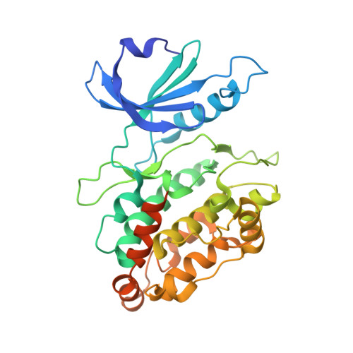
A Pef/Y Substrate Recognition and Signature Motif Plays a Critical Role in Dapk-Related Kinase Activity.

Temmerman, K.De Diego, I.Pogenberg, V.Simon, B.Jonko, W.Li, X.Wilmanns, M.

(2014) Chem Biol 21: 264

  • DOI: https://doi.org/10.1016/j.chembiol.2013.12.008
  • Primary Citation of Related Structures:  
    2W4K, 2XUU, 4B4L

  • PubMed Abstract: 

    Knowledge about protein kinase substrate preferences is biased toward residues immediately adjacent to the site of phosphorylation. By a combined structural, biochemical, and cellular approach, we have discovered an unexpected substrate recognition element with the consensus sequence PEF/Y in the tumor suppressor death-associated protein kinase 1. This motif can be effectively blocked by a specific pseudosubstrate-type interaction with an autoregulatory domain of this kinase. In this arrangement, the central PEF/Y glutamate interacts with a conserved arginine distant to the phosphorylation site in sequence and structure. We also demonstrate that the element is crucial for kinase activity regulation and substrate recognition. The PEF/Y motif distinguishes close death-associated protein kinase relatives from canonical calcium/calmodulin-dependent protein kinases. Insight into this signature and mode of action offers new opportunities to identify specific small molecule inhibitors in PEF/Y-containing protein kinases.


  • Organizational Affiliation

    European Molecular Biology Laboratory Hamburg, Notkestrasse 85, 22603 Hamburg, Germany; European Molecular Biology Laboratory Heidelberg, Meyerhofstrasse 1, 69117 Heidelberg, Germany.


Macromolecules
Find similar proteins by:  (by identity cutoff)  |  3D Structure
Entity ID: 1
MoleculeChains Sequence LengthOrganismDetailsImage
DEATH-ASSOCIATED PROTEIN KINASE 1336Homo sapiensMutation(s): 1 
EC: 2.7.11.1
UniProt & NIH Common Fund Data Resources
Find proteins for P53355 (Homo sapiens)
Explore P53355 
Go to UniProtKB:  P53355
PHAROS:  P53355
GTEx:  ENSG00000196730 
Entity Groups  
Sequence Clusters30% Identity50% Identity70% Identity90% Identity95% Identity100% Identity
UniProt GroupP53355
Sequence Annotations
Expand
  • Reference Sequence
Small Molecules
Ligands 2 Unique
IDChains Name / Formula / InChI Key2D Diagram3D Interactions
1PE
Query on 1PE

Download Ideal Coordinates CCD File 
B [auth A],
C [auth A],
D [auth A]
PENTAETHYLENE GLYCOL
C10 H22 O6
JLFNLZLINWHATN-UHFFFAOYSA-N
SO4
Query on SO4

Download Ideal Coordinates CCD File 
E [auth A]
F [auth A]
G [auth A]
H [auth A]
I [auth A]
E [auth A],
F [auth A],
G [auth A],
H [auth A],
I [auth A],
J [auth A],
K [auth A]
SULFATE ION
O4 S
QAOWNCQODCNURD-UHFFFAOYSA-L
Experimental Data & Validation

Experimental Data

  • Method: X-RAY DIFFRACTION
  • Resolution: 1.75 Å
  • R-Value Free: 0.220 
  • R-Value Work: 0.192 
  • R-Value Observed: 0.193 
  • Space Group: P 21 21 21
Unit Cell:
Length ( Å )Angle ( ˚ )
a = 49.385α = 90
b = 83.798β = 90
c = 84.603γ = 90
Software Package:
Software NamePurpose
REFMACrefinement
XDSdata reduction
SCALAdata scaling
MOLREPphasing

Structure Validation

View Full Validation Report


Entry History 

Deposition Data

Revision History  (Full details and data files)

  • Version 1.0: 2013-08-21
    Type: Initial release
  • Version 1.1: 2014-02-05
    Changes: Database references
  • Version 1.2: 2014-03-05
    Changes: Database references
  • Version 1.3: 2023-12-20
    Changes: Data collection, Database references, Derived calculations, Other, Refinement description