4B30

Structure of the mutant V44A of the fluorescent protein KillerRed


Experimental Data Snapshot

  • Method: X-RAY DIFFRACTION
  • Resolution: 2.10 Å
  • R-Value Free: 0.199 
  • R-Value Work: 0.165 
  • R-Value Observed: 0.167 

wwPDB Validation   3D Report Full Report


This is version 2.1 of the entry. See complete history


Literature

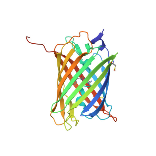
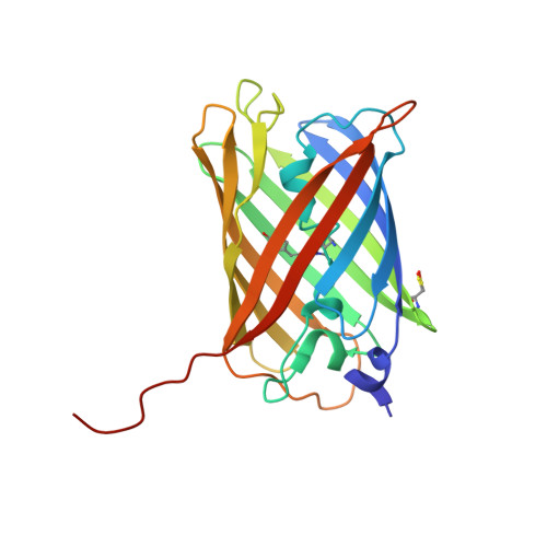
Gfp-Like Phototransformation Mechanisms in the Cytotoxic Fluorescent Protein Killerred Unraveled by Structural and Spectroscopic Investigations.

De Rosny, E.Carpentier, P.

(2012) J Am Chem Soc 134: 18015

  • DOI: https://doi.org/10.1021/ja3073337
  • Primary Citation of Related Structures:  
    4B30

  • PubMed Abstract: 

    KillerRed (KR) is a red fluorescent protein recognized as an efficient genetically encoded photosensitizer. KR generates reactive oxygen species via a complex process of photoreactions, ending up in photobleaching, the mechanism of which remains obscure. In order to clarify these mechanisms, we focus on a single mutant V44A (A44-KR) exhibiting the solely green component of KR. We report on the laser-induced structural transformations of A44-KR at cryogenic temperature, which we have investigated by combining UV-vis fluorescence/absorption spectroscopy with X-ray crystallography. Like the well-known GFP, A44-KR possesses a mixture of protonated (A) absorbing at 397 and deprotonated (B) absorbing at 515 nm chromophores, which are stressed by intense prolonged violet and blue laser sources. Both illuminations directly drive the B-chromophores toward a bleached trans isomerized form. A-type chromophores are sensitive only to violet illumination and are phototransformed either into a deprotonated green fluorescent form by decarboxylation of E218 or into a bleached form with a disordered p-hydroxybenzylidene. In crystallo spectroscopy at cryo-temperature allowed the identification and dissection of an exhaustive scheme of intermediates and end-products resulting from the phototransformation of A44-KR. This constitutes a framework for understanding the photochemistry of the photosensitizer KillerRed.


  • Organizational Affiliation

    Institut de Biologie Structurale Jean-Pierre Ebel, Groupe Métalloprotéines, UMR 5075, Université Joseph Fourier Grenoble 1, CEA, CNRS, 41 rue Horowitz, 38027, Grenoble Cedex 1, France.


Macromolecules
Find similar proteins by:  (by identity cutoff)  |  3D Structure
Entity ID: 1
MoleculeChains Sequence LengthOrganismDetailsImage
KILLERRED235Anthomedusae sp. DC-2005Mutation(s): 2 
UniProt
Find proteins for Q2TCH5 (Anthomedusae sp. DC-2005)
Explore Q2TCH5 
Go to UniProtKB:  Q2TCH5
Entity Groups  
Sequence Clusters30% Identity50% Identity70% Identity90% Identity95% Identity100% Identity
UniProt GroupQ2TCH5
Sequence Annotations
Expand
  • Reference Sequence
Find similar proteins by:  (by identity cutoff)  |  3D Structure
Entity ID: 2
MoleculeChains Sequence LengthOrganismDetailsImage
KILLERRED235Anthomedusae sp. DC-2005Mutation(s): 2 
UniProt
Find proteins for Q2TCH5 (Anthomedusae sp. DC-2005)
Explore Q2TCH5 
Go to UniProtKB:  Q2TCH5
Entity Groups  
Sequence Clusters30% Identity50% Identity70% Identity90% Identity95% Identity100% Identity
UniProt GroupQ2TCH5
Sequence Annotations
Expand
  • Reference Sequence
Small Molecules
Ligands 4 Unique
IDChains Name / Formula / InChI Key2D Diagram3D Interactions
SO4
Query on SO4

Download Ideal Coordinates CCD File 
C [auth A]SULFATE ION
O4 S
QAOWNCQODCNURD-UHFFFAOYSA-L
GOL
Query on GOL

Download Ideal Coordinates CCD File 
K [auth A]
L [auth A]
M [auth A]
N [auth A]
Q [auth B]
K [auth A],
L [auth A],
M [auth A],
N [auth A],
Q [auth B],
R [auth B]
GLYCEROL
C3 H8 O3
PEDCQBHIVMGVHV-UHFFFAOYSA-N
CL
Query on CL

Download Ideal Coordinates CCD File 
D [auth A]
E [auth A]
F [auth A]
G [auth A]
J [auth A]
D [auth A],
E [auth A],
F [auth A],
G [auth A],
J [auth A],
O [auth B],
P [auth B],
S [auth B]
CHLORIDE ION
Cl
VEXZGXHMUGYJMC-UHFFFAOYSA-M
NA
Query on NA

Download Ideal Coordinates CCD File 
H [auth A],
I [auth A]
SODIUM ION
Na
FKNQFGJONOIPTF-UHFFFAOYSA-N
Modified Residues  2 Unique
IDChains TypeFormula2D DiagramParent
CRQ
Query on CRQ
A
L-PEPTIDE LINKINGC16 H16 N4 O5GLN, TYR, GLY
CSO
Query on CSO
A
L-PEPTIDE LINKINGC3 H7 N O3 SCYS
Experimental Data & Validation

Experimental Data

  • Method: X-RAY DIFFRACTION
  • Resolution: 2.10 Å
  • R-Value Free: 0.199 
  • R-Value Work: 0.165 
  • R-Value Observed: 0.167 
  • Space Group: P 62
Unit Cell:
Length ( Å )Angle ( ˚ )
a = 134.31α = 90
b = 134.31β = 90
c = 76.22γ = 120
Software Package:
Software NamePurpose
REFMACrefinement
XDSdata reduction
XSCALEdata scaling
PHASERphasing

Structure Validation

View Full Validation Report


Entry History 

Deposition Data

Revision History  (Full details and data files)

  • Version 1.0: 2013-01-16
    Type: Initial release
  • Version 1.1: 2019-05-08
    Changes: Data collection, Derived calculations, Experimental preparation, Other
  • Version 1.2: 2019-10-23
    Changes: Data collection, Database references, Other
  • Version 2.0: 2023-11-15
    Changes: Atomic model, Data collection, Database references, Derived calculations
  • Version 2.1: 2023-12-20
    Changes: Refinement description