4AYG

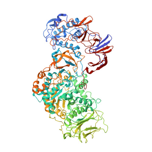
Lactobacillus reuteri N-terminally truncated glucansucrase GTF180 in orthorhombic apo-form


Experimental Data Snapshot

  • Method: X-RAY DIFFRACTION
  • Resolution: 2.00 Å
  • R-Value Free: 0.183 
  • R-Value Work: 0.155 
  • R-Value Observed: 0.156 

wwPDB Validation   3D Report Full Report


This is version 1.4 of the entry. See complete history


Literature

Flexibility of Truncated and Full-Length Glucansucrase Gtf180 Enzymes from Lactobacillus Reuteri 180.

Pijning, T.Vujicic-Zagar, A.Kralj, S.Dijkhuizen, L.Dijkstra, B.W.

(2014) FEBS J 281: 2159

  • DOI: https://doi.org/10.1111/febs.12769
  • Primary Citation of Related Structures:  
    4AYG

  • PubMed Abstract: 

    Glucansucrase enzymes synthesize high-molecular-mass extracellular α-glucan polysaccharides from sucrose. Previously, the crystal structure of truncated glucansucrase glucosyltransferase (GTF)180-ΔN from Lactobacillus reuteri 180 (lacking the N-terminal domain) revealed an elongated overall structure with two remote domains (IV and V) extending away from the core. By contrast, a new crystal form of the α-1,6/α-1,3 specific glucansucrase GTF180-ΔN shows an approximate 120(o) rotation of domain V about a hinge located between domains IV and V, giving a much more compact structure than before. Positional variability of domain V in solution is confirmed by small angle X-ray scattering experiments and rigid-body ensemble calculations. In addition, small angle X-ray scattering measurements of full-length GTF180 also provide the first structural data for a full-length glucansucrase, showing that the enzyme has an almost symmetric boomerang-like molecular shape, with a bend likely located between domains IV and V. The ~ 700-residue N-terminal domain, which is not present in the crystal structures, extends away from domain V and the catalytic core of the enzyme. We conclude that, as a result of the hinge region, in solution, GTF180-ΔN (and likely also the full-length GTF180) shows conformational flexibility; this may be a general feature of GH70 glucansucrases.


  • Organizational Affiliation

    Laboratory of Biophysical Chemistry, Groningen Biomolecular Sciences and Biotechnology Institute (GBB), University of Groningen, The Netherlands.


Macromolecules
Find similar proteins by:  (by identity cutoff)  |  3D Structure
Entity ID: 1
MoleculeChains Sequence LengthOrganismDetailsImage
GLUCANSUCRASE
A, B
1,039Limosilactobacillus reuteriMutation(s): 0 
EC: 2.4.1.5
UniProt
Find proteins for Q5SBN3 (Limosilactobacillus reuteri)
Explore Q5SBN3 
Go to UniProtKB:  Q5SBN3
Entity Groups  
Sequence Clusters30% Identity50% Identity70% Identity90% Identity95% Identity100% Identity
UniProt GroupQ5SBN3
Sequence Annotations
Expand
  • Reference Sequence
Small Molecules
Ligands 5 Unique
IDChains Name / Formula / InChI Key2D Diagram3D Interactions
SO4
Query on SO4

Download Ideal Coordinates CCD File 
BC [auth B]
CC [auth B]
DB [auth A]
DC [auth B]
EC [auth B]
BC [auth B],
CC [auth B],
DB [auth A],
DC [auth B],
EC [auth B],
FC [auth B],
GC [auth B],
HC [auth B],
IC [auth B],
JC [auth B],
KA [auth A],
KC [auth B],
LA [auth A],
MA [auth A],
NA [auth A],
OA [auth A],
PA [auth A],
QA [auth A],
RA [auth A],
SA [auth A],
TA [auth A]
SULFATE ION
O4 S
QAOWNCQODCNURD-UHFFFAOYSA-L
GOL
Query on GOL

Download Ideal Coordinates CCD File 
AA [auth A]
AC [auth B]
BA [auth A]
CA [auth A]
D [auth A]
AA [auth A],
AC [auth B],
BA [auth A],
CA [auth A],
D [auth A],
DA [auth A],
E [auth A],
EA [auth A],
F [auth A],
FA [auth A],
FB [auth B],
G [auth A],
GA [auth A],
GB [auth B],
H [auth A],
HA [auth A],
HB [auth B],
I [auth A],
IA [auth A],
IB [auth B],
J [auth A],
JA [auth A],
JB [auth B],
K [auth A],
KB [auth B],
L [auth A],
LB [auth B],
M [auth A],
MB [auth B],
N [auth A],
NB [auth B],
O [auth A],
OB [auth B],
P [auth A],
PB [auth B],
Q [auth A],
QB [auth B],
R [auth A],
RB [auth B],
S [auth A],
SB [auth B],
T [auth A],
TB [auth B],
U [auth A],
UB [auth B],
V [auth A],
VB [auth B],
W [auth A],
WB [auth B],
X [auth A],
XB [auth B],
Y [auth A],
YB [auth B],
Z [auth A],
ZB [auth B]
GLYCEROL
C3 H8 O3
PEDCQBHIVMGVHV-UHFFFAOYSA-N
IPA
Query on IPA

Download Ideal Coordinates CCD File 
CB [auth A],
QC [auth B]
ISOPROPYL ALCOHOL
C3 H8 O
KFZMGEQAYNKOFK-UHFFFAOYSA-N
ACY
Query on ACY

Download Ideal Coordinates CCD File 
AB [auth A]
BB [auth A]
LC [auth B]
MC [auth B]
NC [auth B]
AB [auth A],
BB [auth A],
LC [auth B],
MC [auth B],
NC [auth B],
OC [auth B],
PC [auth B],
UA [auth A],
VA [auth A],
WA [auth A],
XA [auth A],
YA [auth A],
ZA [auth A]
ACETIC ACID
C2 H4 O2
QTBSBXVTEAMEQO-UHFFFAOYSA-N
CA
Query on CA

Download Ideal Coordinates CCD File 
C [auth A],
EB [auth B]
CALCIUM ION
Ca
BHPQYMZQTOCNFJ-UHFFFAOYSA-N
Experimental Data & Validation

Experimental Data

  • Method: X-RAY DIFFRACTION
  • Resolution: 2.00 Å
  • R-Value Free: 0.183 
  • R-Value Work: 0.155 
  • R-Value Observed: 0.156 
  • Space Group: P 21 21 21
Unit Cell:
Length ( Å )Angle ( ˚ )
a = 92.792α = 90
b = 147.363β = 90
c = 244.374γ = 90
Software Package:
Software NamePurpose
REFMACrefinement
HKL-2000data reduction
HKLdata scaling
PHASERphasing

Structure Validation

View Full Validation Report


Entry History 

Deposition Data

Revision History  (Full details and data files)

  • Version 1.0: 2013-07-03
    Type: Initial release
  • Version 1.1: 2014-03-19
    Changes: Database references
  • Version 1.2: 2014-05-14
    Changes: Database references
  • Version 1.3: 2019-07-17
    Changes: Data collection
  • Version 1.4: 2023-12-20
    Changes: Data collection, Database references, Derived calculations, Other, Refinement description