Crystal Direct: A New Method for Automated Crystal Harvesting Based on Laser-Induced Photoablation of Thin Films
Cipriani, F., Rower, M., Landret, C., Zander, U., Felisaz, F., Marquez, J.A.(2012) Acta Crystallogr D Biol Crystallogr 68: 1393
- PubMed: 22993093
- DOI: https://doi.org/10.1107/S0907444912031459
- Primary Citation of Related Structures:
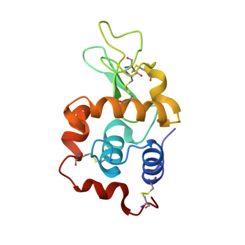
4AXR, 4AXT, 4AXU, 4B0D - PubMed Abstract:
The use of automated systems for crystallization and X-ray data collection is now widespread. However, these two steps are separated by the need to transfer crystals from crystallization supports to X-ray data-collection supports, which is a difficult manual operation. Here, a new approach is proposed called CrystalDirect (CD) which enables full automation of the crystal-harvesting process. In this approach, crystals are grown on ultrathin films in a newly designed vapour-diffusion crystallization plate and are recovered by excision of the film through laser-induced photoablation. The film pieces containing crystals are then directly attached to a pin for X-ray data collection. This new method eliminates the delicate step of `crystal fishing', thereby enabling full automation of the crystal-mounting process. Additional advantages of this approach include the absence of mechanical stress and that it facilitates handling of microcrystals. The CD crystallization plates are also suitable for in situ crystal screening with minimal X-ray background. This method could enable the operational integration of highly automated crystallization and data-collection facilities, minimizing the delay between crystal identification and diffraction measurements. It can also contribute significantly to the advancement of challenging projects that require the systematic testing of large numbers of crystals.
Organizational Affiliation:
Grenoble Outstation, European Molecular Biology Laboratory, 6 Rue Jules Horowitz, 38042 Grenoble, France. cipriani@embl.fr