4AUB

the complex Structure of the bacterial aldo-keto reductase AKR14A1 with NADP and citrate


Experimental Data Snapshot

  • Method: X-RAY DIFFRACTION
  • Resolution: 2.05 Å
  • R-Value Free: 0.238 
  • R-Value Work: 0.202 
  • R-Value Observed: 0.203 

wwPDB Validation   3D Report Full Report


Ligand Structure Quality Assessment 


This is version 1.2 of the entry. See complete history


Literature

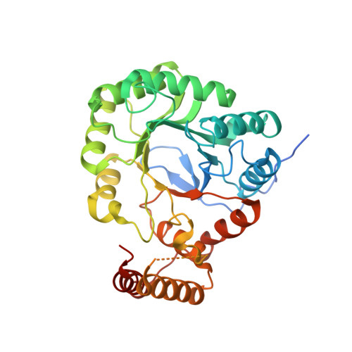
The Diversity of Microbial Aldo/Keto Reductases from Escherichia Coli K12.

Lapthorn, A.J.Zhu, X.Ellis, E.M.

(2013) Chem Biol Interact 202: 168

  • DOI: https://doi.org/10.1016/j.cbi.2012.10.008
  • Primary Citation of Related Structures:  
    4AST, 4AUB

  • PubMed Abstract: 

    The genome of Escherichia coli K12 contains 9 open reading frames encoding aldo/keto reductases (AKRs) that are differentially regulated and sequence diverse. A significant amount of data is available for the E. coli AKRs through the availability of gene knockouts and gene expression studies, which adds to the biochemical and kinetic data. This together with the availability of crystal structures for nearly half of the E. coli AKRs and homologues of several others provides an opportunity to look at the diversity of these representative bacterial AKRs. Based around the common AKR fold of (β/α)8 barrel with two additional α-helices, the E. coli AKRs have a loop structure that is more diverse than their mammalian counterparts, creating a variety of active site architectures. Nearly half of the AKRs are expected to be monomeric, but there are examples of dimeric, trimeric and octameric enzymes, as well as diversity in specificity for NAD as well as NADP as a cofactor. However in functional assignments and characterisation of enzyme activities there is a paucity of data when compared to the mammalian AKR enzymes.


  • Organizational Affiliation

    School of Chemistry, University of Glasgow, Glasgow, United Kingdom. adrian.lapthorn@glasgow.ac.uk


Macromolecules
Find similar proteins by:  (by identity cutoff)  |  3D Structure
Entity ID: 1
MoleculeChains Sequence LengthOrganismDetailsImage
ALDO-KETO REDUCTASE AKR14A1
A, B, C, D, E
A, B, C, D, E, F, G, H
366Escherichia coli K-12Mutation(s): 0 
UniProt
Find proteins for Q46851 (Escherichia coli (strain K12))
Explore Q46851 
Go to UniProtKB:  Q46851
Entity Groups  
Sequence Clusters30% Identity50% Identity70% Identity90% Identity95% Identity100% Identity
UniProt GroupQ46851
Sequence Annotations
Expand
  • Reference Sequence
Small Molecules
Ligands 2 Unique
IDChains Name / Formula / InChI Key2D Diagram3D Interactions
NAP
Query on NAP

Download Ideal Coordinates CCD File 
I [auth A]
K [auth B]
M [auth C]
P [auth E]
R [auth F]
I [auth A],
K [auth B],
M [auth C],
P [auth E],
R [auth F],
T [auth G],
V [auth H]
NADP NICOTINAMIDE-ADENINE-DINUCLEOTIDE PHOSPHATE
C21 H28 N7 O17 P3
XJLXINKUBYWONI-NNYOXOHSSA-N
FLC
Query on FLC

Download Ideal Coordinates CCD File 
J [auth A]
L [auth B]
N [auth C]
O [auth D]
Q [auth E]
J [auth A],
L [auth B],
N [auth C],
O [auth D],
Q [auth E],
S [auth F],
U [auth G],
W [auth H]
CITRATE ANION
C6 H5 O7
KRKNYBCHXYNGOX-UHFFFAOYSA-K
Experimental Data & Validation

Experimental Data

  • Method: X-RAY DIFFRACTION
  • Resolution: 2.05 Å
  • R-Value Free: 0.238 
  • R-Value Work: 0.202 
  • R-Value Observed: 0.203 
  • Space Group: P 1 21 1
Unit Cell:
Length ( Å )Angle ( ˚ )
a = 91.813α = 90
b = 191.092β = 105.97
c = 97.303γ = 90
Software Package:
Software NamePurpose
REFMACrefinement
HKLdata reduction
SCALEPACKdata scaling
AMoREphasing

Structure Validation

View Full Validation Report


Ligand Structure Quality Assessment 


Entry History 

Deposition Data

Revision History  (Full details and data files)

  • Version 1.0: 2012-06-06
    Type: Initial release
  • Version 1.1: 2014-03-12
    Changes: Database references, Source and taxonomy
  • Version 1.2: 2023-12-20
    Changes: Data collection, Database references, Derived calculations, Other, Refinement description