4ATJ

DISTAL HEME POCKET MUTANT (H42E) OF RECOMBINANT HORSERADISH PEROXIDASE IN COMPLEX WITH BENZHYDROXAMIC ACID


Experimental Data Snapshot

  • Method: X-RAY DIFFRACTION
  • Resolution: 2.50 Å
  • R-Value Free: 0.194 
  • R-Value Work: 0.162 

wwPDB Validation   3D Report Full Report


Ligand Structure Quality Assessment 


This is version 1.5 of the entry. See complete history


Literature

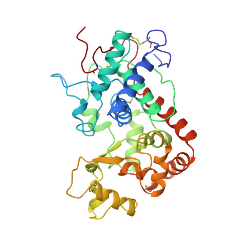
Structural analysis of the two horseradish peroxidase catalytic residue variants H42E and R38S/H42E: implications for the catalytic cycle.

Meno, K.Jennings, S.Smith, A.T.Henriksen, A.Gajhede, M.

(2002) Acta Crystallogr D Biol Crystallogr 58: 1803-1812

  • DOI: https://doi.org/10.1107/s090744490201329x
  • Primary Citation of Related Structures:  
    1KZM, 4ATJ

  • PubMed Abstract: 

    The crystal structures of horseradish peroxidase C (HRPC) active-site mutants H42E and R38S/H42E co-crystallized with benzhydroxamic acid (BHA) and ferulic acid (FA), respectively, have been solved. The 2.5 A crystal structure of the H42E-BHA complex reveals that the side-chain O atoms of Glu42 occupy positions that are very similar to the positions of the two side-chain N atoms of the distal histidine in the wild-type HRPC-BHA structure. The mutation disturbs the hydrogen-bonding network extending from residue 42 to the distal calcium ion and results in the absence of the water molecule that is usually ligated to this ion in plant peroxidases. Consequently, the distal calcium ion is six- rather than seven-coordinated. In the 2.0 A R38S/H42E structure the position of Glu42 is different and no FA is observed in the distal haem pocket. This is a consequence of the absence of the Arg38 side chain, which limits the flexibility of the Glu42 side chain and modulates its acidity, making it unsuitable as a general acid-base catalyst in the reaction cycle. The water ligated to the distal calcium ion is present, showing that the wild-type distal hydrogen-bonding network is preserved. These results show why a glutamic acid residue can substitute for the conserved distal histidine in HRPC and that Arg38 plays a significant role in controlling the positioning and ionization state of the residue at position 42. Furthermore, these structures indicate that changes in the distal cavity are conveyed through the distal hydrogen-bonding network to the distal calcium site.


  • Organizational Affiliation

    Protein Structure Group, Department of Chemistry, University of Copenhagen, Universitetsparken 5, DK-2100 Copenhagen Ø, Denmark.


Macromolecules
Find similar proteins by:  (by identity cutoff)  |  3D Structure
Entity ID: 1
MoleculeChains Sequence LengthOrganismDetailsImage
PROTEIN (PEROXIDASE C1A)
A, B
309Armoracia rusticanaMutation(s): 1 
EC: 1.11.1.7
UniProt
Find proteins for P00433 (Armoracia rusticana)
Explore P00433 
Go to UniProtKB:  P00433
Entity Groups  
Sequence Clusters30% Identity50% Identity70% Identity90% Identity95% Identity100% Identity
UniProt GroupP00433
Sequence Annotations
Expand
  • Reference Sequence
Small Molecules
Ligands 3 Unique
IDChains Name / Formula / InChI Key2D Diagram3D Interactions
HEM
Query on HEM

Download Ideal Coordinates CCD File 
E [auth A],
I [auth B]
PROTOPORPHYRIN IX CONTAINING FE
C34 H32 Fe N4 O4
KABFMIBPWCXCRK-RGGAHWMASA-L
BHO
Query on BHO

Download Ideal Coordinates CCD File 
F [auth A],
J [auth B]
BENZHYDROXAMIC ACID
C7 H7 N O2
VDEUYMSGMPQMIK-UHFFFAOYSA-N
CA
Query on CA

Download Ideal Coordinates CCD File 
C [auth A],
D [auth A],
G [auth B],
H [auth B]
CALCIUM ION
Ca
BHPQYMZQTOCNFJ-UHFFFAOYSA-N
Experimental Data & Validation

Experimental Data

  • Method: X-RAY DIFFRACTION
  • Resolution: 2.50 Å
  • R-Value Free: 0.194 
  • R-Value Work: 0.162 
  • Space Group: P 1 21 1
Unit Cell:
Length ( Å )Angle ( ˚ )
a = 74.93α = 90
b = 62.27β = 104.27
c = 78.23γ = 90
Software Package:
Software NamePurpose
DENZOdata reduction
SCALEPACKdata scaling
AMoREphasing
X-PLORrefinement

Structure Validation

View Full Validation Report


Ligand Structure Quality Assessment 


Entry History 

Deposition Data

Revision History  (Full details and data files)

  • Version 1.0: 2002-10-02
    Type: Initial release
  • Version 1.1: 2007-10-16
    Changes: Version format compliance
  • Version 1.2: 2011-07-13
    Changes: Version format compliance
  • Version 1.3: 2018-03-14
    Changes: Database references
  • Version 1.4: 2019-11-27
    Changes: Database references, Derived calculations
  • Version 1.5: 2023-09-13
    Changes: Data collection, Database references, Derived calculations, Refinement description