4ACL

3D Structure of DotU from Francisella novicida


Experimental Data Snapshot

  • Method: X-RAY DIFFRACTION
  • Resolution: 2.49 Å
  • R-Value Free: 0.279 
  • R-Value Work: 0.218 
  • R-Value Observed: 0.221 

wwPDB Validation   3D Report Full Report


This is version 1.3 of the entry. See complete history


Literature

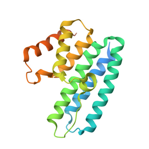
The Structure of the Conserved Type Six Secretion Protein Tssl (Dotu) from Francisella Novicida

Robb, C.S.Nano, F.E.Boraston, A.B.

(2012) J Mol Biol 419: 277

  • DOI: https://doi.org/10.1016/j.jmb.2012.04.003
  • Primary Citation of Related Structures:  
    4ACK, 4ACL

  • PubMed Abstract: 

    Type six secretion systems (T6SSs) are found in many Gram-negative bacteria and are important for their virulence or their ecological competitiveness. The multicomponent T6SSs are responsible for the translocation of effector molecules into target eukaryotic or prokaryotic cells. The Francisella pathogenicity island encodes a putative T6SS that Francisella novicida requires for intramacrophage growth and virulence during infection of rodents. Here, we present the X-ray crystal structure of the conserved type six secretion component TssL (DotU) from F. novicida. The structure of this protein, which is referred to as Ftn_TssL, revealed an all-α-helical fold that is a unique fusion of two 3-helix bundles. The sequence of Ftn_TssL shows low identity to presumed homologs that are found in most T6SSs. The structure of Ftn_TssL, however, has allowed us to provide bioinformatics evidence that the F. novicida TssL has a fold that is very likely representative for TssL forms from both T6SSs and from the distantly related B subclass of type four secretion systems. A map of sequence conservation on the TssL structure revealed a surface-exposed groove that may represent a functional site on the protein.


  • Organizational Affiliation

    Department of Biochemistry and Microbiology, University of Victoria, PO Box 3055 Station CSC, Victoria, British Columbia, Canada V8W 3P6.


Macromolecules
Find similar proteins by:  (by identity cutoff)  |  3D Structure
Entity ID: 1
MoleculeChains Sequence LengthOrganismDetailsImage
TSSL
A, B
205Francisella tularensis subsp. novicida U112Mutation(s): 0 
UniProt
Find proteins for A0Q7H7 (Francisella tularensis subsp. novicida (strain U112))
Explore A0Q7H7 
Go to UniProtKB:  A0Q7H7
Entity Groups  
Sequence Clusters30% Identity50% Identity70% Identity90% Identity95% Identity100% Identity
UniProt GroupA0Q7H7
Sequence Annotations
Expand
  • Reference Sequence
Small Molecules
Ligands 3 Unique
IDChains Name / Formula / InChI Key2D Diagram3D Interactions
AU
Query on AU

Download Ideal Coordinates CCD File 
F [auth A]
G [auth A]
H [auth A]
I [auth A]
J [auth A]
F [auth A],
G [auth A],
H [auth A],
I [auth A],
J [auth A],
K [auth A],
L [auth B],
M [auth B],
N [auth B],
O [auth B],
P [auth B],
Q [auth B]
GOLD ION
Au
ZBKIUFWVEIBQRT-UHFFFAOYSA-N
EDO
Query on EDO

Download Ideal Coordinates CCD File 
D [auth A],
E [auth A]
1,2-ETHANEDIOL
C2 H6 O2
LYCAIKOWRPUZTN-UHFFFAOYSA-N
NA
Query on NA

Download Ideal Coordinates CCD File 
C [auth A]SODIUM ION
Na
FKNQFGJONOIPTF-UHFFFAOYSA-N
Experimental Data & Validation

Experimental Data

  • Method: X-RAY DIFFRACTION
  • Resolution: 2.49 Å
  • R-Value Free: 0.279 
  • R-Value Work: 0.218 
  • R-Value Observed: 0.221 
  • Space Group: P 1 21 1
Unit Cell:
Length ( Å )Angle ( ˚ )
a = 39.309α = 90
b = 85.314β = 102.38
c = 66.989γ = 90
Software Package:
Software NamePurpose
REFMACrefinement
d*TREKdata reduction
d*TREKdata scaling
autoSHARPphasing

Structure Validation

View Full Validation Report


Entry History 

Deposition Data

Revision History  (Full details and data files)

  • Version 1.0: 2012-04-25
    Type: Initial release
  • Version 1.1: 2012-06-06
    Changes: Other
  • Version 1.2: 2017-07-05
    Changes: Data collection
  • Version 1.3: 2017-09-13
    Changes: Data collection