4AAO

MacA-H93G


Experimental Data Snapshot

  • Method: X-RAY DIFFRACTION
  • Resolution: 2.30 Å
  • R-Value Free: 0.241 
  • R-Value Work: 0.190 
  • R-Value Observed: 0.192 

wwPDB Validation   3D Report Full Report


Ligand Structure Quality Assessment 


This is version 1.1 of the entry. See complete history


Literature

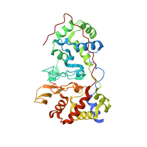
Maca is a Second Cytochrome C Peroxidase of Geobacter Sulfurreducens.

Seidel, J.Hoffmann, M.Ellis, K.E.Seidel, A.Spatzal, T.Gerhardt, S.Elliott, S.J.Einsle, O.

(2012) Biochemistry 51: 2747

  • DOI: https://doi.org/10.1021/bi300249u
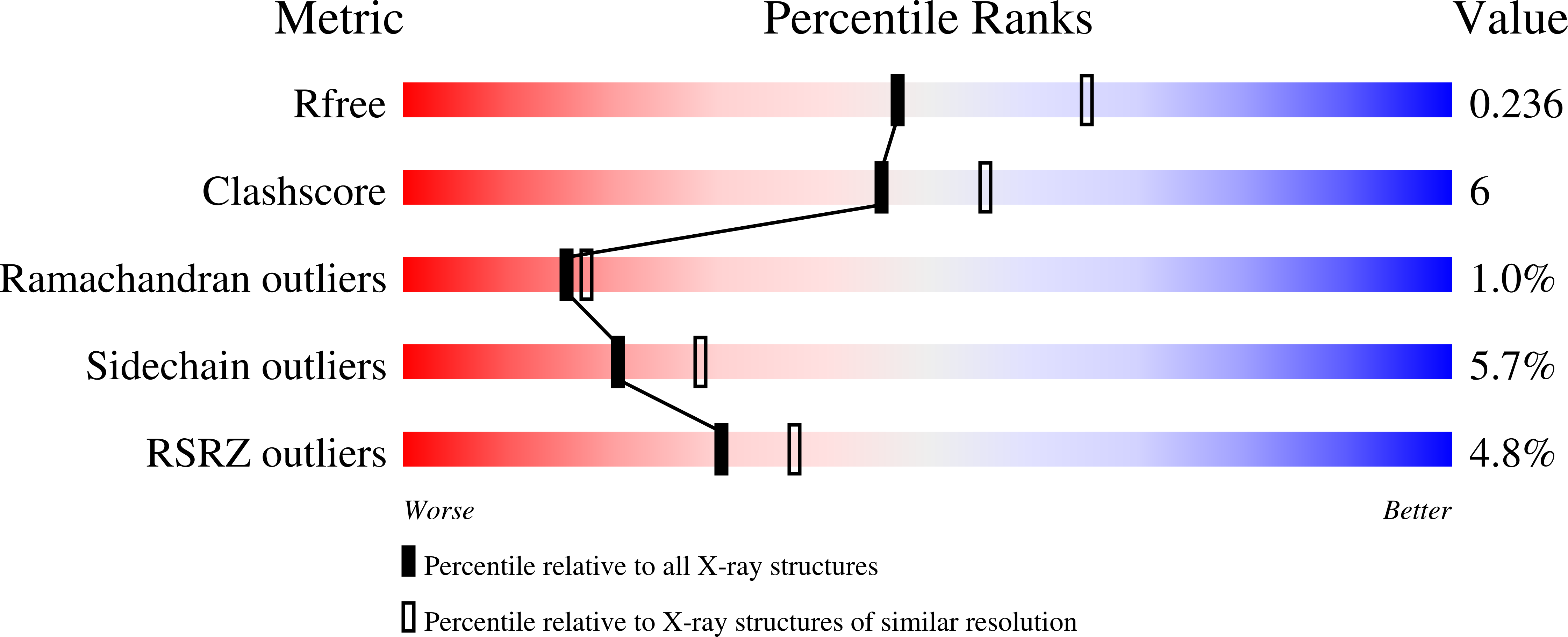
  • Primary Citation of Related Structures:  
    4AAL, 4AAM, 4AAN, 4AAO

  • PubMed Abstract: 

    The metal-reducing δ-proteobacterium Geobacter sulfurreducens produces a large number of c-type cytochromes, many of which have been implicated in the transfer of electrons to insoluble metal oxides. Among these, the dihemic MacA was assigned a central role. Here we have produced G. sulfurreducens MacA by recombinant expression in Escherichia coli and have solved its three-dimensional structure in three different oxidation states. Sequence comparisons group MacA into the family of diheme cytochrome c peroxidases, and the protein indeed showed hydrogen peroxide reductase activity with ABTS(-2) as an electron donor. The observed K(M) was 38.5 ± 3.7 μM H(2)O(2) and v(max) was 0.78 ± 0.03 μmol of H(2)O(2)·min(-1)·mg(-1), resulting in a turnover number k(cat) = 0.46 · s(-1). In contrast, no Fe(III) reductase activity was observed. MacA was found to display electrochemical properties similar to other bacterial diheme peroxidases, in addition to the ability to electrochemically mediate electron transfer to the soluble cytochrome PpcA. Differences in activity between CcpA and MacA can be rationalized with structural variations in one of the three loop regions, loop 2, that undergoes conformational changes during reductive activation of the enzyme. This loop is adjacent to the active site heme and forms an open loop structure rather than a more rigid helix as in CcpA. For the activation of the protein, the loop has to displace the distal ligand to the active site heme, H93, in loop 1. A H93G variant showed an unexpected formation of a helix in loop 2 and disorder in loop 1, while a M297H variant that altered the properties of the electron transfer heme abolished reductive activation.


  • Organizational Affiliation

    Lehrstuhl für Biochemie, Institut für Organische Chemie und Biochemie, Albert-Ludwigs-Universität Freiburg, Albertstraße 21, 79104 Freiburg, Germany.


Macromolecules
Find similar proteins by:  (by identity cutoff)  |  3D Structure
Entity ID: 1
MoleculeChains Sequence LengthOrganismDetailsImage
CYTOCHROME C551 PEROXIDASE
A, B
341Geobacter sulfurreducens PCAMutation(s): 1 
EC: 1.11.1.5
UniProt
Find proteins for Q74FY6 (Geobacter sulfurreducens (strain ATCC 51573 / DSM 12127 / PCA))
Explore Q74FY6 
Go to UniProtKB:  Q74FY6
Entity Groups  
Sequence Clusters30% Identity50% Identity70% Identity90% Identity95% Identity100% Identity
UniProt GroupQ74FY6
Sequence Annotations
Expand
  • Reference Sequence
Small Molecules
Ligands 3 Unique
IDChains Name / Formula / InChI Key2D Diagram3D Interactions
HEC
Query on HEC

Download Ideal Coordinates CCD File 
C [auth A],
D [auth A],
L [auth B],
M [auth B]
HEME C
C34 H34 Fe N4 O4
HXQIYSLZKNYNMH-LJNAALQVSA-N
SO4
Query on SO4

Download Ideal Coordinates CCD File 
F [auth A]
G [auth A]
H [auth A]
I [auth A]
J [auth A]
F [auth A],
G [auth A],
H [auth A],
I [auth A],
J [auth A],
K [auth A],
O [auth B],
P [auth B],
Q [auth B],
R [auth B],
S [auth B]
SULFATE ION
O4 S
QAOWNCQODCNURD-UHFFFAOYSA-L
CA
Query on CA

Download Ideal Coordinates CCD File 
E [auth A],
N [auth B]
CALCIUM ION
Ca
BHPQYMZQTOCNFJ-UHFFFAOYSA-N
Experimental Data & Validation

Experimental Data

  • Method: X-RAY DIFFRACTION
  • Resolution: 2.30 Å
  • R-Value Free: 0.241 
  • R-Value Work: 0.190 
  • R-Value Observed: 0.192 
  • Space Group: P 65 2 2
Unit Cell:
Length ( Å )Angle ( ˚ )
a = 118.719α = 90
b = 118.719β = 90
c = 245.98γ = 120
Software Package:
Software NamePurpose
REFMACrefinement
iMOSFLMdata reduction
SCALAdata scaling
MOLREPphasing

Structure Validation

View Full Validation Report


Ligand Structure Quality Assessment 


Entry History 

Deposition Data

  • Released Date: 2012-10-17 
  • Deposition Author(s): Seidel, J.

Revision History  (Full details and data files)

  • Version 1.0: 2012-10-17
    Type: Initial release
  • Version 1.1: 2023-12-20
    Changes: Data collection, Database references, Derived calculations, Other, Refinement description