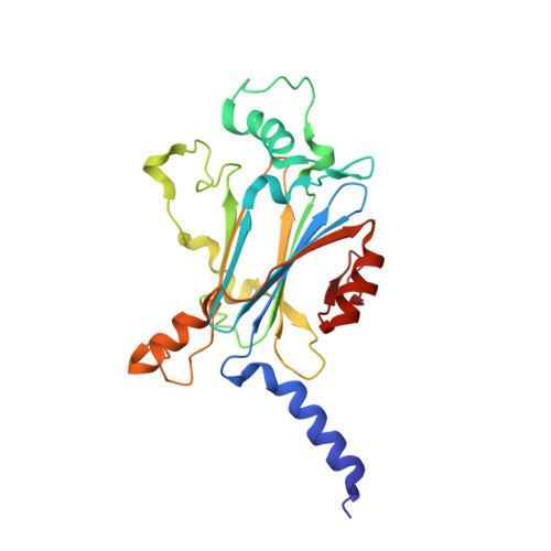
4ZQX

A revised partiality model and post-refinement algorithm for X-ray free-electron laser data


Experimental Data Snapshot

  • Method: X-RAY DIFFRACTION
  • Resolution: 1.46 Å
  • R-Value Free: 0.153 
  • R-Value Work: 0.108 
  • R-Value Observed: 0.110 

wwPDB Validation   3D Report Full Report


Ligand Structure Quality Assessment 


This is version 1.5 of the entry. See complete history


Literature

A revised partiality model and post-refinement algorithm for X-ray free-electron laser data.

Ginn, H.M.Brewster, A.S.Hattne, J.Evans, G.Wagner, A.Grimes, J.M.Sauter, N.K.Sutton, G.Stuart, D.I.

(2015) Acta Crystallogr D Biol Crystallogr 71: 1400-1410

  • DOI: https://doi.org/10.1107/S1399004715006902
  • Primary Citation of Related Structures:  
    4ZQX

  • PubMed Abstract: 

    Research towards using X-ray free-electron laser (XFEL) data to solve structures using experimental phasing methods such as sulfur single-wavelength anomalous dispersion (SAD) has been hampered by shortcomings in the diffraction models for X-ray diffraction from FELs. Owing to errors in the orientation matrix and overly simple partiality models, researchers have required large numbers of images to converge to reliable estimates for the structure-factor amplitudes, which may not be feasible for all biological systems. Here, data for cytoplasmic polyhedrosis virus type 17 (CPV17) collected at 1.3 Å wavelength at the Linac Coherent Light Source (LCLS) are revisited. A previously published definition of a partiality model for reflections illuminated by self-amplified spontaneous emission (SASE) pulses is built upon, which defines a fraction between 0 and 1 based on the intersection of a reflection with a spread of Ewald spheres modelled by a super-Gaussian wavelength distribution in the X-ray beam. A method of post-refinement to refine the parameters of this model is suggested. This has generated a merged data set with an overall discrepancy (by calculating the R(split) value) of 3.15% to 1.46 Å resolution from a 7225-image data set. The atomic numbers of C, N and O atoms in the structure are distinguishable in the electron-density map. There are 13 S atoms within the 237 residues of CPV17, excluding the initial disordered methionine. These only possess 0.42 anomalous scattering electrons each at 1.3 Å wavelength, but the 12 that have single predominant positions are easily detectable in the anomalous difference Fourier map. It is hoped that these improvements will lead towards XFEL experimental phase determination and structure determination by sulfur SAD and will generally increase the utility of the method for difficult cases.


  • Organizational Affiliation

    Division of Structural Biology, Wellcome Trust Centre for Human Genetics, Roosevelt Drive, Oxford OX3 7BN, England.


Macromolecules
Find similar proteins by:  (by identity cutoff)  |  3D Structure
Entity ID: 1
MoleculeChains Sequence LengthOrganismDetailsImage
Polyhedrin237Uranotaenia sapphirina cypovirusMutation(s): 0 
UniProt
Find proteins for Q5EK29 (Uranotaenia sapphirina cypovirus)
Explore Q5EK29 
Go to UniProtKB:  Q5EK29
Entity Groups  
Sequence Clusters30% Identity50% Identity70% Identity90% Identity95% Identity100% Identity
UniProt GroupQ5EK29
Sequence Annotations
Expand
  • Reference Sequence
Small Molecules
Ligands 2 Unique
IDChains Name / Formula / InChI Key2D Diagram3D Interactions
ATP
Query on ATP

Download Ideal Coordinates CCD File 
B [auth A]ADENOSINE-5'-TRIPHOSPHATE
C10 H16 N5 O13 P3
ZKHQWZAMYRWXGA-KQYNXXCUSA-N
MG
Query on MG

Download Ideal Coordinates CCD File 
C [auth A]MAGNESIUM ION
Mg
JLVVSXFLKOJNIY-UHFFFAOYSA-N
Experimental Data & Validation

Experimental Data

  • Method: X-RAY DIFFRACTION
  • Resolution: 1.46 Å
  • R-Value Free: 0.153 
  • R-Value Work: 0.108 
  • R-Value Observed: 0.110 
  • Space Group: I 2 3
Unit Cell:
Length ( Å )Angle ( ˚ )
a = 106.1α = 90
b = 106.1β = 90
c = 106.1γ = 90
Software Package:
Software NamePurpose
PHENIXrefinement
cctbx.xfeldata reduction
MOLREPphasing

Structure Validation

View Full Validation Report


Ligand Structure Quality Assessment 


Entry History & Funding Information

Deposition Data


Funding OrganizationLocationGrant Number
Medical Research Council (United Kingdom)United KingdomG1000099
Wellcome TrustUnited Kingdom090532/Z/09/Z
Wellcome TrustUnited Kingdom075491/04
National Institutes of HealthUnited KingdomGM095887
National Institutes of HealthUnited KingdomGM102520

Revision History  (Full details and data files)

  • Version 1.0: 2015-06-03
    Type: Initial release
  • Version 1.1: 2015-06-24
    Changes: Database references
  • Version 1.2: 2017-09-13
    Changes: Author supporting evidence
  • Version 1.3: 2018-01-17
    Changes: Data collection
  • Version 1.4: 2018-11-14
    Changes: Data collection
  • Version 1.5: 2024-01-10
    Changes: Data collection, Database references, Derived calculations, Refinement description