4ZP2

Crystal structure of E. coli multidrug transporter MdfA in complex with n-dodecyl-N,N-dimethylamine-N-oxide


Experimental Data Snapshot

  • Method: X-RAY DIFFRACTION
  • Resolution: 2.20 Å
  • R-Value Free: 0.232 
  • R-Value Work: 0.206 
  • R-Value Observed: 0.208 

wwPDB Validation   3D Report Full Report


Ligand Structure Quality Assessment 


This is version 1.2 of the entry. See complete history


Literature

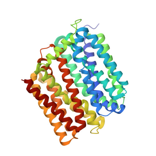
Substrate-bound structure of the E. coli multidrug resistance transporter MdfA

Heng, J.Zhao, Y.Liu, M.Liu, Y.Fan, J.Wang, X.Zhao, Y.Zhang, X.C.

(2015) Cell Res 25: 1060-1073

  • DOI: https://doi.org/10.1038/cr.2015.94
  • Primary Citation of Related Structures:  
    4ZOW, 4ZP0, 4ZP2

  • PubMed Abstract: 

    Multidrug resistance is a serious threat to public health. Proton motive force-driven antiporters from the major facilitator superfamily (MFS) constitute a major group of multidrug-resistance transporters. Currently, no reports on crystal structures of MFS antiporters in complex with their substrates exist. The E. coli MdfA transporter is a well-studied model system for biochemical analyses of multidrug-resistance MFS antiporters. Here, we report three crystal structures of MdfA-ligand complexes at resolutions up to 2.0 Å, all in the inward-facing conformation. The substrate-binding site sits proximal to the conserved acidic residue, D34. Our mutagenesis studies support the structural observations of the substrate-binding mode and the notion that D34 responds to substrate binding by adjusting its protonation status. Taken together, our data unveil the substrate-binding mode of MFS antiporters and suggest a mechanism of transport via this group of transporters.


  • Organizational Affiliation

    National Laboratory of Macromolecules, National Center of Protein Science-Beijing, Institute of Biophysics, Chinese Academy of Sciences, 15 Datun Road, Beijing 100101, China.


Macromolecules
Find similar proteins by:  (by identity cutoff)  |  3D Structure
Entity ID: 1
MoleculeChains Sequence LengthOrganismDetailsImage
Multidrug transporter MdfA392Escherichia coli K-12Mutation(s): 1 
Gene Names: mdfAcmlAcmrb0842JW0826
Membrane Entity: Yes 
UniProt
Find proteins for P0AEY8 (Escherichia coli (strain K12))
Explore P0AEY8 
Go to UniProtKB:  P0AEY8
Entity Groups  
Sequence Clusters30% Identity50% Identity70% Identity90% Identity95% Identity100% Identity
UniProt GroupP0AEY8
Sequence Annotations
Expand
  • Reference Sequence
Small Molecules
Experimental Data & Validation

Experimental Data

  • Method: X-RAY DIFFRACTION
  • Resolution: 2.20 Å
  • R-Value Free: 0.232 
  • R-Value Work: 0.206 
  • R-Value Observed: 0.208 
  • Space Group: C 1 2 1
Unit Cell:
Length ( Å )Angle ( ˚ )
a = 94.086α = 90
b = 64.085β = 104.19
c = 101.253γ = 90
Software Package:
Software NamePurpose
SCALEPACKdata scaling
PHENIXrefinement
PDB_EXTRACTdata extraction
DENZOdata reduction
SOLVEphasing

Structure Validation

View Full Validation Report


Ligand Structure Quality Assessment 


Entry History 

Deposition Data

Revision History  (Full details and data files)

  • Version 1.0: 2015-08-19
    Type: Initial release
  • Version 1.1: 2015-09-16
    Changes: Database references
  • Version 1.2: 2024-03-20
    Changes: Data collection, Database references, Derived calculations