4ZGH

Structure of Sugar Binding Protein Pneumolysin


Experimental Data Snapshot

  • Method: X-RAY DIFFRACTION
  • Resolution: 2.90 Å
  • R-Value Free: 0.307 
  • R-Value Work: 0.199 
  • R-Value Observed: 0.204 

wwPDB Validation   3D Report Full Report


This is version 1.3 of the entry. See complete history


Literature

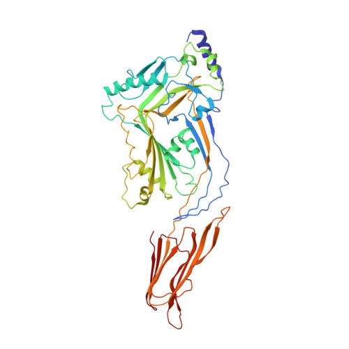
Crystal structure of Streptococcus pneumoniae pneumolysin provides key insights into early steps of pore formation.

Lawrence, S.L.Feil, S.C.Morton, C.J.Farrand, A.J.Mulhern, T.D.Gorman, M.A.Wade, K.R.Tweten, R.K.Parker, M.W.

(2015) Sci Rep 5: 14352-14352

  • DOI: https://doi.org/10.1038/srep14352
  • Primary Citation of Related Structures:  
    4ZGH

  • PubMed Abstract: 

    Pore-forming proteins are weapons often used by bacterial pathogens to breach the membrane barrier of target cells. Despite their critical role in infection important structural aspects of the mechanism of how these proteins assemble into pores remain unknown. Streptococcus pneumoniae is the world's leading cause of pneumonia, meningitis, bacteremia and otitis media. Pneumolysin (PLY) is a major virulence factor of S. pneumoniae and a target for both small molecule drug development and vaccines. PLY is a member of the cholesterol-dependent cytolysins (CDCs), a family of pore-forming toxins that form gigantic pores in cell membranes. Here we present the structure of PLY determined by X-ray crystallography and, in solution, by small-angle X-ray scattering. The crystal structure reveals PLY assembles as a linear oligomer that provides key structural insights into the poorly understood early monomer-monomer interactions of CDCs at the membrane surface.


  • Organizational Affiliation

    ACRF Rational Drug Discovery Centre, St. Vincent's Institute of Medical Research, Fitzroy, Victoria 3065, Australia.


Macromolecules
Find similar proteins by:  (by identity cutoff)  |  3D Structure
Entity ID: 1
MoleculeChains Sequence LengthOrganismDetailsImage
Pneumolysin477Streptococcus pneumoniae D39Mutation(s): 3 
Gene Names: plySPD_1726
Membrane Entity: Yes 
UniProt
Find proteins for Q04IN8 (Streptococcus pneumoniae serotype 2 (strain D39 / NCTC 7466))
Explore Q04IN8 
Go to UniProtKB:  Q04IN8
Entity Groups  
Sequence Clusters30% Identity50% Identity70% Identity90% Identity95% Identity100% Identity
UniProt GroupQ04IN8
Sequence Annotations
Expand
  • Reference Sequence
Small Molecules
Ligands 3 Unique
IDChains Name / Formula / InChI Key2D Diagram3D Interactions
AUC
Query on AUC

Download Ideal Coordinates CCD File 
I [auth A],
J [auth A]
GOLD (I) CYANIDE ION
C2 Au N2
DGOHFTDNMSZWDQ-UHFFFAOYSA-N
PEG
Query on PEG

Download Ideal Coordinates CCD File 
G [auth A],
H [auth A]
DI(HYDROXYETHYL)ETHER
C4 H10 O3
MTHSVFCYNBDYFN-UHFFFAOYSA-N
EDO
Query on EDO

Download Ideal Coordinates CCD File 
B [auth A],
C [auth A],
D [auth A],
E [auth A],
F [auth A]
1,2-ETHANEDIOL
C2 H6 O2
LYCAIKOWRPUZTN-UHFFFAOYSA-N
Experimental Data & Validation

Experimental Data

  • Method: X-RAY DIFFRACTION
  • Resolution: 2.90 Å
  • R-Value Free: 0.307 
  • R-Value Work: 0.199 
  • R-Value Observed: 0.204 
  • Space Group: P 21 21 21
Unit Cell:
Length ( Å )Angle ( ˚ )
a = 24.857α = 90
b = 133.619β = 90
c = 220.442γ = 90
Software Package:
Software NamePurpose
PHENIXrefinement
Aimlessdata scaling
PHASERphasing
XDSdata scaling

Structure Validation

View Full Validation Report


Entry History 

Deposition Data

Revision History  (Full details and data files)

  • Version 1.0: 2016-03-09
    Type: Initial release
  • Version 1.1: 2017-11-22
    Changes: Advisory, Data collection, Derived calculations, Refinement description
  • Version 1.2: 2019-02-13
    Changes: Data collection, Source and taxonomy
  • Version 1.3: 2023-09-27
    Changes: Data collection, Database references, Refinement description