4YL3

Crystal Structures of mPGES-1 Inhibitor Complexes


Experimental Data Snapshot

  • Method: X-RAY DIFFRACTION
  • Resolution: 1.41 Å
  • R-Value Free: 0.183 
  • R-Value Work: 0.166 
  • R-Value Observed: 0.167 

wwPDB Validation   3D Report Full Report


Ligand Structure Quality Assessment 


This is version 1.2 of the entry. See complete history


Literature

Crystal Structures of mPGES-1 Inhibitor Complexes Form a Basis for the Rational Design of Potent Analgesic and Anti-Inflammatory Therapeutics.

Luz, J.G.Antonysamy, S.Kuklish, S.L.Condon, B.Lee, M.R.Allison, D.Yu, X.P.Chandrasekhar, S.Backer, R.Zhang, A.Russell, M.Chang, S.S.Harvey, A.Sloan, A.V.Fisher, M.J.

(2015) J Med Chem 58: 4727-4737

  • DOI: https://doi.org/10.1021/acs.jmedchem.5b00330
  • Primary Citation of Related Structures:  
    4YK5, 4YL0, 4YL1, 4YL3

  • PubMed Abstract: 

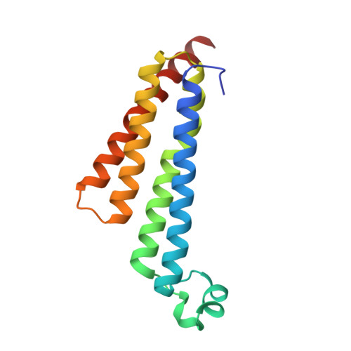
    Microsomal prostaglandin E synthase 1 (mPGES-1) is an α-helical homotrimeric integral membrane inducible enzyme that catalyzes the formation of prostaglandin E2 (PGE2) from prostaglandin H2 (PGH2). Inhibition of mPGES-1 has been proposed as a therapeutic strategy for the treatment of pain, inflammation, and some cancers. Interest in mPGES-1 inhibition can, in part, be attributed to the potential circumvention of cardiovascular risks associated with anti-inflammatory cyclooxygenase 2 inhibitors (coxibs) by targeting the prostaglandin pathway downstream of PGH2 synthesis and avoiding suppression of antithrombotic prostacyclin production. We determined the crystal structure of mPGES-1 bound to four potent inhibitors in order to understand their structure-activity relationships and provide a framework for the rational design of improved molecules. In addition, we developed a light-scattering-based thermal stability assay to identify molecules for crystallographic studies.


  • Organizational Affiliation

    †Lilly Biotechnology Center San Diego, 10300 Campus Point Drive, Suite 200, San Diego, California 92121, United States.


Macromolecules
Find similar proteins by:  (by identity cutoff)  |  3D Structure
Entity ID: 1
MoleculeChains Sequence LengthOrganismDetailsImage
Prostaglandin E synthase148Homo sapiensMutation(s): 0 
Gene Names: PTGESMGST1L1MPGES1PGESPIG12
EC: 5.3.99.3
Membrane Entity: Yes 
UniProt & NIH Common Fund Data Resources
Find proteins for O14684 (Homo sapiens)
Explore O14684 
Go to UniProtKB:  O14684
PHAROS:  O14684
GTEx:  ENSG00000148344 
Entity Groups  
Sequence Clusters30% Identity50% Identity70% Identity90% Identity95% Identity100% Identity
UniProt GroupO14684
Sequence Annotations
Expand
  • Reference Sequence
Small Molecules
Binding Affinity Annotations 
IDSourceBinding Affinity
4U9 Binding MOAD:  4YL3 IC50: 3940 (nM) from 1 assay(s)
Experimental Data & Validation

Experimental Data

  • Method: X-RAY DIFFRACTION
  • Resolution: 1.41 Å
  • R-Value Free: 0.183 
  • R-Value Work: 0.166 
  • R-Value Observed: 0.167 
  • Space Group: H 3
Unit Cell:
Length ( Å )Angle ( ˚ )
a = 76.434α = 90
b = 76.434β = 90
c = 123.264γ = 120
Software Package:
Software NamePurpose
REFMACrefinement
PDB_EXTRACTdata extraction
Cootmodel building
SCALAdata scaling
SHELXphasing

Structure Validation

View Full Validation Report


Ligand Structure Quality Assessment 


Entry History 

Deposition Data

Revision History  (Full details and data files)

  • Version 1.0: 2015-07-08
    Type: Initial release
  • Version 1.1: 2020-07-29
    Type: Remediation
    Reason: Carbohydrate remediation
    Changes: Data collection, Database references, Derived calculations, Source and taxonomy, Structure summary
  • Version 1.2: 2024-02-28
    Changes: Data collection, Database references, Structure summary