4XQC

Crystal Structure of the Homospermidine Synthase (HSS) from Blastochloris viridis in Complex with NAD and 1,3-diaminopropane.


Experimental Data Snapshot

  • Method: X-RAY DIFFRACTION
  • Resolution: 1.27 Å
  • R-Value Free: 0.182 
  • R-Value Work: 0.150 
  • R-Value Observed: 0.152 

wwPDB Validation   3D Report Full Report


Ligand Structure Quality Assessment 


This is version 1.2 of the entry. See complete history


Literature

Comprehensive Structural Characterization of the Bacterial Homospermidine Synthase-an Essential Enzyme of the Polyamine Metabolism.

Krossa, S.Faust, A.Ober, D.Scheidig, A.J.

(2016) Sci Rep 6: 19501-19501

  • DOI: https://doi.org/10.1038/srep19501
  • Primary Citation of Related Structures:  
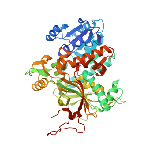
    4PLP, 4TVB, 4XQ9, 4XQC, 4XQE, 4XQG, 4XR4, 4XRG

  • PubMed Abstract: 

    The highly conserved bacterial homospermidine synthase (HSS) is a key enzyme of the polyamine metabolism of many proteobacteria including pathogenic strains such as Legionella pneumophila and Pseudomonas aeruginosa; The unique usage of NAD(H) as a prosthetic group is a common feature of bacterial HSS, eukaryotic HSS and deoxyhypusine synthase (DHS). The structure of the bacterial enzyme does not possess a lysine residue in the active center and thus does not form an enzyme-substrate Schiff base intermediate as observed for the DHS. In contrast to the DHS the active site is not formed by the interface of two subunits but resides within one subunit of the bacterial HSS. Crystal structures of Blastochloris viridis HSS (BvHSS) reveal two distinct substrate binding sites, one of which is highly specific for putrescine. BvHSS features a side pocket in the direct vicinity of the active site formed by conserved amino acids and a potential substrate discrimination, guiding, and sensing mechanism. The proposed reaction steps for the catalysis of BvHSS emphasize cation-π interaction through a conserved Trp residue as a key stabilizer of high energetic transition states.


  • Organizational Affiliation

    Structural Biology-Zoological Institute, Kiel University, Am Botanischen Garten 11, 24118 Kiel, Germany.


Macromolecules
Find similar proteins by:  (by identity cutoff)  |  3D Structure
Entity ID: 1
MoleculeChains Sequence LengthOrganismDetailsImage
Homospermidine synthase
A, B
475Blastochloris viridisMutation(s): 0 
Gene Names: hss
EC: 2.5.1.44
UniProt
Find proteins for O32323 (Blastochloris viridis)
Explore O32323 
Go to UniProtKB:  O32323
Entity Groups  
Sequence Clusters30% Identity50% Identity70% Identity90% Identity95% Identity100% Identity
UniProt GroupO32323
Sequence Annotations
Expand
  • Reference Sequence
Small Molecules
Ligands 4 Unique
IDChains Name / Formula / InChI Key2D Diagram3D Interactions
NAD
Query on NAD

Download Ideal Coordinates CCD File 
C [auth A],
J [auth B]
NICOTINAMIDE-ADENINE-DINUCLEOTIDE
C21 H27 N7 O14 P2
BAWFJGJZGIEFAR-NNYOXOHSSA-N
1PS
Query on 1PS

Download Ideal Coordinates CCD File 
F [auth A],
K [auth B]
3-PYRIDINIUM-1-YLPROPANE-1-SULFONATE
C8 H11 N O3 S
REEBJQTUIJTGAL-UHFFFAOYSA-N
13D
Query on 13D

Download Ideal Coordinates CCD File 
D [auth A],
E [auth A]
1,3-DIAMINOPROPANE
C3 H10 N2
XFNJVJPLKCPIBV-UHFFFAOYSA-N
ACT
Query on ACT

Download Ideal Coordinates CCD File 
G [auth A],
H [auth A],
I [auth A],
L [auth B]
ACETATE ION
C2 H3 O2
QTBSBXVTEAMEQO-UHFFFAOYSA-M
Experimental Data & Validation

Experimental Data

  • Method: X-RAY DIFFRACTION
  • Resolution: 1.27 Å
  • R-Value Free: 0.182 
  • R-Value Work: 0.150 
  • R-Value Observed: 0.152 
  • Space Group: P 2 21 21
Unit Cell:
Length ( Å )Angle ( ˚ )
a = 60.075α = 90
b = 109.791β = 90
c = 157.342γ = 90
Software Package:
Software NamePurpose
PHENIXrefinement
XDSdata reduction
SCALAdata scaling
MOLREPphasing
PDB_EXTRACTdata extraction

Structure Validation

View Full Validation Report


Ligand Structure Quality Assessment 


Entry History 

Deposition Data

  • Released Date: 2016-01-27 
  • Deposition Author(s): Krossa, S.

Revision History  (Full details and data files)

  • Version 1.0: 2016-01-27
    Type: Initial release
  • Version 1.1: 2018-03-07
    Changes: Data collection
  • Version 1.2: 2024-01-10
    Changes: Data collection, Database references, Refinement description, Structure summary