4XDW

Crystal Structure of the L74F/M78V/I80V/L114F mutant of LEH


Experimental Data Snapshot

  • Method: X-RAY DIFFRACTION
  • Resolution: 2.05 Å
  • R-Value Free: 0.245 
  • R-Value Work: 0.202 
  • R-Value Observed: 0.204 

wwPDB Validation   3D Report Full Report


This is version 1.2 of the entry. See complete history


Literature

Reshaping an Enzyme Binding Pocket for Enhanced and Inverted Stereoselectivity: Use of Smallest Amino Acid Alphabets in Directed Evolution

Sun, Z.Lonsdale, R.Kong, X.D.Xu, J.H.Zhou, J.Reetz, M.T.

(2015) Angew Chem Int Ed Engl 54: 12410-12415

  • DOI: https://doi.org/10.1002/anie.201501809
  • Primary Citation of Related Structures:  
    4XBT, 4XBX, 4XBY, 4XDV, 4XDW

  • PubMed Abstract: 

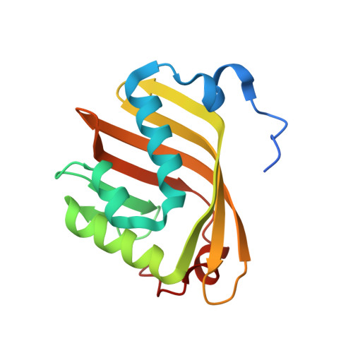
    Directed evolution based on saturation mutagenesis at sites lining the binding pocket is a commonly practiced strategy for enhancing or inverting the stereoselectivity of enzymes for use in organic chemistry or biotechnology. However, as the number of residues in a randomization site increases to five or more, the screening effort for 95 % library coverage increases astronomically until it is no longer feasible. We propose the use of a single amino acid for saturation mutagenesis at superlarge randomization sites comprising 10 or more residues. When used to reshape the binding pocket of limonene epoxide hydrolase, this strategy, which drastically reduces the search space and thus the screening effort, resulted in R,R- and S,S-selective mutants for the hydrolytic desymmetrization of cyclohexene oxide and other epoxides. X-ray crystal structures and docking studies of the mutants unveiled the source of stereoselectivity and shed light on the mechanistic intricacies of this enzyme.


  • Organizational Affiliation

    Max-Planck-Institut für Kohlenforschung, Kaiser-Wilhelm-Platz 1, 45470 Mülheim an der Ruhr (Germany).


Macromolecules
Find similar proteins by:  (by identity cutoff)  |  3D Structure
Entity ID: 1
MoleculeChains Sequence LengthOrganismDetailsImage
Limonene-1,2-epoxide hydrolase155Rhodococcus erythropolisMutation(s): 4 
Gene Names: limA
EC: 3.3.2.8
UniProt
Find proteins for Q9ZAG3 (Rhodococcus erythropolis)
Explore Q9ZAG3 
Go to UniProtKB:  Q9ZAG3
Entity Groups  
Sequence Clusters30% Identity50% Identity70% Identity90% Identity95% Identity100% Identity
UniProt GroupQ9ZAG3
Sequence Annotations
Expand
  • Reference Sequence
Experimental Data & Validation

Experimental Data

  • Method: X-RAY DIFFRACTION
  • Resolution: 2.05 Å
  • R-Value Free: 0.245 
  • R-Value Work: 0.202 
  • R-Value Observed: 0.204 
  • Space Group: P 1 21 1
Unit Cell:
Length ( Å )Angle ( ˚ )
a = 104.267α = 90
b = 60.901β = 90.57
c = 119.05γ = 90
Software Package:
Software NamePurpose
HKL-2000data reduction
PHASERphasing
PHENIXrefinement
PDB_EXTRACTdata extraction
HKL-2000data scaling
Cootmodel building

Structure Validation

View Full Validation Report


Entry History 

Deposition Data

Revision History  (Full details and data files)

  • Version 1.0: 2015-07-15
    Type: Initial release
  • Version 1.1: 2015-10-14
    Changes: Database references
  • Version 1.2: 2023-11-08
    Changes: Data collection, Database references, Derived calculations, Refinement description