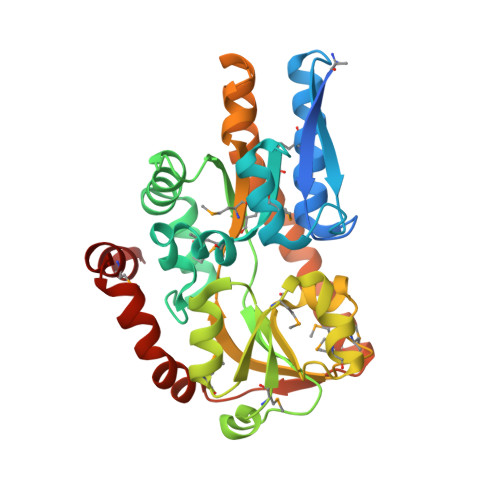
CRYSTAL STRUCTURE OF A TRAP PERIPLASMIC SOLUTE BINDING PROTEIN FROM CITROBACTER KOSERI (CKO_04899, TARGET EFI-510094) WITH BOUND D-glucuronate
Yadava, U., Vetting, M.W., Al Obaidi, N.F., Toro, R., Morisco, L.L., Benach, J., Wasserman, S.R., Attonito, J.D., Scott Glenn, A., Chamala, S., Chowdhury, S., Lafleur, J., Love, J., Seidel, R.D., Whalen, K.L., Gerlt, J.A., Almo, S.C., Enzyme Function Initiative (EFI)To be published.