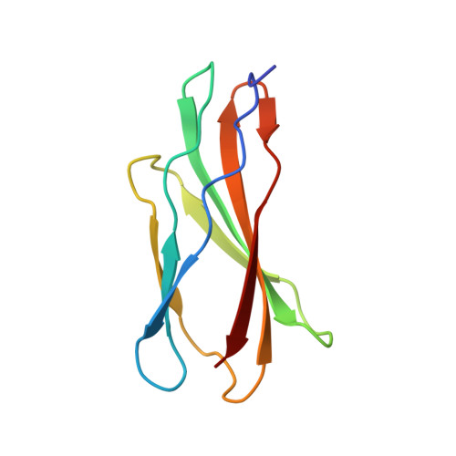
4WTW

Crystal structure of the third FnIII domain of integrin beta4


Experimental Data Snapshot

  • Method: X-RAY DIFFRACTION
  • Resolution: 1.61 Å
  • R-Value Free: 0.180 
  • R-Value Work: 0.151 
  • R-Value Observed: 0.152 

wwPDB Validation   3D Report Full Report


This is version 2.0 of the entry. See complete history


Literature

Combination of X-ray crystallography, SAXS and DEER to obtain the structure of the FnIII-3,4 domains of integrin alpha6beta4

Alonso-Garcia, N.Garcia-Rubio, I.Manso, J.A.Buey, R.M.Urien, H.Sonnenberg, A.Jeschke, G.de Pereda, J.M.

(2015) Acta Crystallogr D Biol Crystallogr 71: 969-985

  • DOI: https://doi.org/10.1107/S1399004715002485
  • Primary Citation of Related Structures:  
    4WTW, 4WTX

  • PubMed Abstract: 

    Integrin α6β4 is a major component of hemidesmosomes that mediate the stable anchorage of epithelial cells to the underlying basement membrane. Integrin α6β4 has also been implicated in cell proliferation and migration and in carcinoma progression. The third and fourth fibronectin type III domains (FnIII-3,4) of integrin β4 mediate binding to the hemidesmosomal proteins BPAG1e and BPAG2, and participate in signalling. Here, it is demonstrated that X-ray crystallography, small-angle X-ray scattering and double electron-electron resonance (DEER) complement each other to solve the structure of the FnIII-3,4 region. The crystal structures of the individual FnIII-3 and FnIII-4 domains were solved and the relative arrangement of the FnIII domains was elucidated by combining DEER with site-directed spin labelling. Multiple structures of the interdomain linker were modelled by Monte Carlo methods complying with DEER constraints, and the final structures were selected against experimental scattering data. FnIII-3,4 has a compact and cambered flat structure with an evolutionary conserved surface that is likely to correspond to a protein-interaction site. Finally, this hybrid method is of general application for the study of other macromolecules and complexes.


  • Organizational Affiliation

    Instituto de Biología Molecular y Celular del Cancer, Consejo Superior de Investigaciones Científicas - University of Salamanca, Campus Unamuno, 37007 Salamanca, Spain.


Macromolecules
Find similar proteins by:  (by identity cutoff)  |  3D Structure
Entity ID: 1
MoleculeChains Sequence LengthOrganismDetailsImage
Integrin beta-4
A, B
96Homo sapiensMutation(s): 0 
Gene Names: ITGB4
UniProt & NIH Common Fund Data Resources
Find proteins for P16144 (Homo sapiens)
Explore P16144 
Go to UniProtKB:  P16144
PHAROS:  P16144
GTEx:  ENSG00000132470 
Entity Groups  
Sequence Clusters30% Identity50% Identity70% Identity90% Identity95% Identity100% Identity
UniProt GroupP16144
Sequence Annotations
Expand
  • Reference Sequence
Experimental Data & Validation

Experimental Data

  • Method: X-RAY DIFFRACTION
  • Resolution: 1.61 Å
  • R-Value Free: 0.180 
  • R-Value Work: 0.151 
  • R-Value Observed: 0.152 
  • Space Group: I 21 21 21
Unit Cell:
Length ( Å )Angle ( ˚ )
a = 66.76α = 90
b = 68.37β = 90
c = 88.49γ = 90
Software Package:
Software NamePurpose
XDSdata reduction
XSCALEdata scaling
PHENIXrefinement
PDB_EXTRACTdata extraction
PHASERphasing

Structure Validation

View Full Validation Report


Entry History & Funding Information

Deposition Data


Funding OrganizationLocationGrant Number
Spanish Ministry of Economy and Competitiveness and the European Regional Development FundSpainBFU2009-08389
Spanish Ministry of Economy and Competitiveness and the European Regional Development FundSpainBFU2012-32847

Revision History  (Full details and data files)

  • Version 1.0: 2015-02-11
    Type: Initial release
  • Version 1.1: 2015-04-08
    Changes: Database references
  • Version 1.2: 2015-04-22
    Changes: Database references
  • Version 2.0: 2024-01-10
    Changes: Atomic model, Data collection, Database references, Refinement description