Capture and X-ray diffraction studies of protein microcrystals in a microfluidic trap array.
Lyubimov, A.Y., Murray, T.D., Koehl, A., Araci, I.E., Uervirojnangkoorn, M., Zeldin, O.B., Cohen, A.E., Soltis, S.M., Baxter, E.L., Brewster, A.S., Sauter, N.K., Brunger, A.T., Berger, J.M.(2015) Acta Crystallogr D Biol Crystallogr 71: 928-940
- PubMed: 25849403
- DOI: https://doi.org/10.1107/S1399004715002308
- Primary Citation of Related Structures:
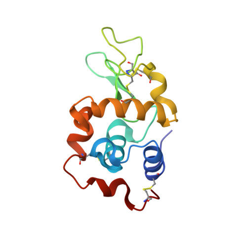
4WMG - PubMed Abstract:
X-ray free-electron lasers (XFELs) promise to enable the collection of interpretable diffraction data from samples that are refractory to data collection at synchrotron sources. At present, however, more efficient sample-delivery methods that minimize the consumption of microcrystalline material are needed to allow the application of XFEL sources to a wide range of challenging structural targets of biological importance. Here, a microfluidic chip is presented in which microcrystals can be captured at fixed, addressable points in a trap array from a small volume (<10 µl) of a pre-existing slurry grown off-chip. The device can be mounted on a standard goniostat for conducting diffraction experiments at room temperature without the need for flash-cooling. Proof-of-principle tests with a model system (hen egg-white lysozyme) demonstrated the high efficiency of the microfluidic approach for crystal harvesting, permitting the collection of sufficient data from only 265 single-crystal still images to permit determination and refinement of the structure of the protein. This work shows that microfluidic capture devices can be readily used to facilitate data collection from protein microcrystals grown in traditional laboratory formats, enabling analysis when cryopreservation is problematic or when only small numbers of crystals are available. Such microfluidic capture devices may also be useful for data collection at synchrotron sources.
Organizational Affiliation:
Department of Molecular and Cellular Physiology, Stanford University, Stanford, CA 94305, USA.