4WKJ

Crystallographic Structure of a Dodecameric RNA-DNA Hybrid


Experimental Data Snapshot

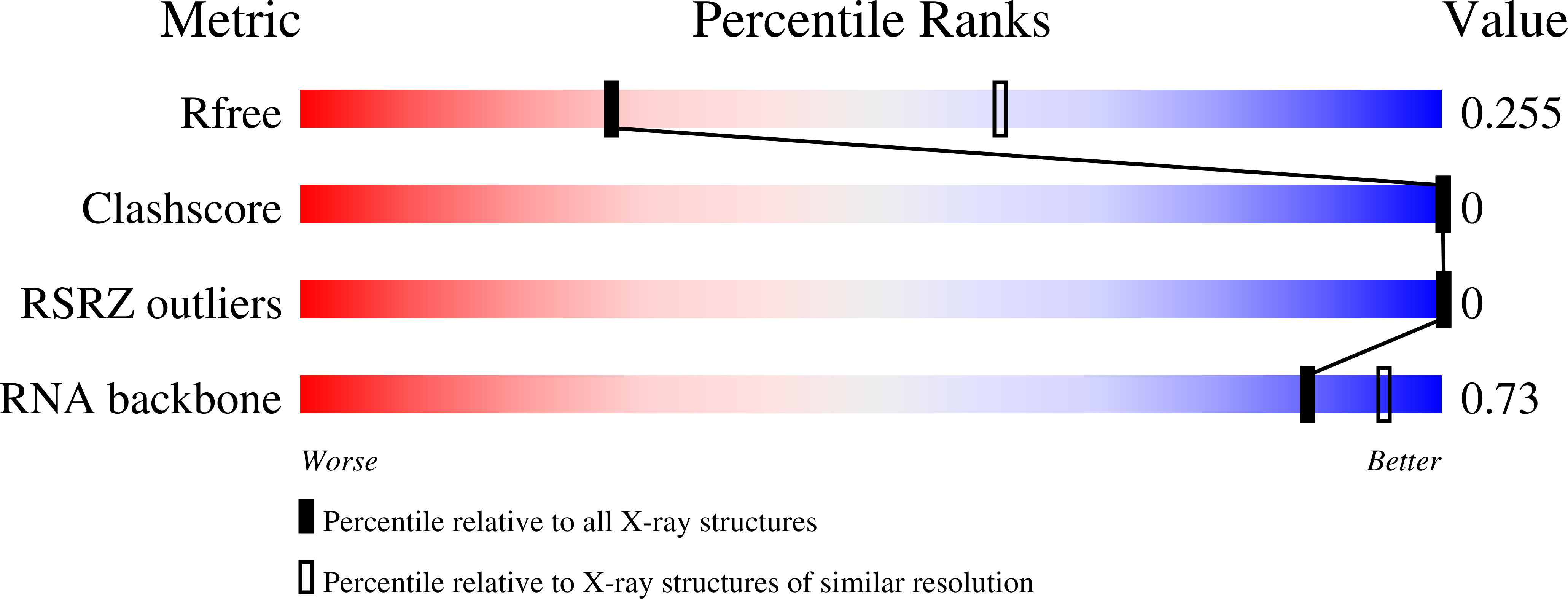
  • Method: X-RAY DIFFRACTION
  • Resolution: 2.80 Å
  • R-Value Free: 0.242 
  • R-Value Work: 0.238 
  • R-Value Observed: 0.239 

wwPDB Validation   3D Report Full Report


This is version 1.5 of the entry. See complete history


Literature

Crystal structure of RNA-DNA duplex provides insight into conformational changes induced by RNase H binding.

Davis, R.R.Shaban, N.M.Perrino, F.W.Hollis, T.

(2015) Cell Cycle 14: 668-673

  • DOI: https://doi.org/10.4161/15384101.2014.994996
  • Primary Citation of Related Structures:  
    4WKJ

  • PubMed Abstract: 

    RNA-DNA hybrids play essential roles in a variety of biological processes, including DNA replication, transcription, and viral integration. Ribonucleotides incorporated within DNA are hydrolyzed by RNase H enzymes in a removal process that is necessary for maintaining genomic stability. In order to understand the structural determinants involved in recognition of a hybrid substrate by RNase H we have determined the crystal structure of a dodecameric non-polypurine/polypyrimidine tract RNA-DNA duplex. A comparison to the same sequence bound to RNase H, reveals structural changes to the duplex that include widening of the major groove to 12.5 Å from 4.2 Å and decreasing the degree of bending along the axis which may play a crucial role in the ribonucleotide recognition and cleavage mechanism within RNase H. This structure allows a direct comparison to be made about the conformational changes induced in RNA-DNA hybrids upon binding to RNase H and may provide insight into how dysfunction in the endonuclease causes disease.


  • Organizational Affiliation

    a Department of Biochemistry; Center for Structural Biology ; Wake Forest School of Medicine ; Winston-Salem , NC USA.


Macromolecules

Find similar nucleic acids by:  Sequence   |   3D Structure  

Entity ID: 1
MoleculeChains LengthOrganismImage
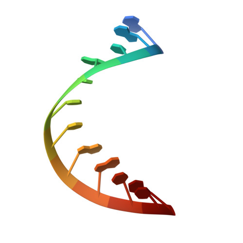
RNA (5'-R(*GP*AP*CP*AP*CP*CP*UP*GP*AP*UP*UP*C)-3')A [auth D],
C [auth A],
E [auth C],
G
12Escherichia coli
Sequence Annotations
Expand
  • Reference Sequence

Find similar nucleic acids by:  Sequence   |   3D Structure  

Entity ID: 2
MoleculeChains LengthOrganismImage
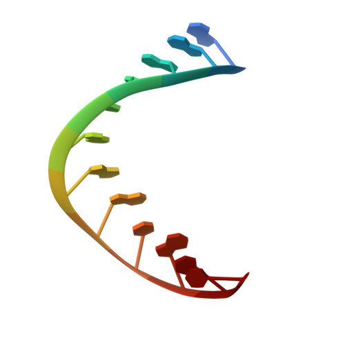
DNA (5'-D(*GP*AP*AP*TP*CP*AP*GP*GP*TP*GP*TP*C)-3')B [auth E],
D [auth B],
F,
H
12Escherichia coli
Sequence Annotations
Expand
  • Reference Sequence
Small Molecules
Ligands 1 Unique
IDChains Name / Formula / InChI Key2D Diagram3D Interactions
MG
Query on MG

Download Ideal Coordinates CCD File 
I [auth F]MAGNESIUM ION
Mg
JLVVSXFLKOJNIY-UHFFFAOYSA-N
Experimental Data & Validation

Experimental Data

  • Method: X-RAY DIFFRACTION
  • Resolution: 2.80 Å
  • R-Value Free: 0.242 
  • R-Value Work: 0.238 
  • R-Value Observed: 0.239 
  • Space Group: P 1 21 1
Unit Cell:
Length ( Å )Angle ( ˚ )
a = 46.649α = 90
b = 44.459β = 105.27
c = 83.346γ = 90
Software Package:
Software NamePurpose
REFMACrefinement
Cootmodel building
CrystalCleardata collection
d*TREKdata scaling
PHASERphasing

Structure Validation

View Full Validation Report


Entry History & Funding Information

Deposition Data


Funding OrganizationLocationGrant Number
American Heart AssociationUnited States10GRNT3650033
National Institutes of Health/National Institute of General Medical Sciences (NIH/NIGMS)United StatesGM069962
National Institutes of Health/National Institute of General Medical Sciences (NIH/NIGMS)United StatesGM108827

Revision History  (Full details and data files)

  • Version 1.0: 2014-10-22
    Type: Initial release
  • Version 1.1: 2014-10-29
    Changes: Derived calculations
  • Version 1.2: 2015-03-04
    Changes: Database references
  • Version 1.3: 2017-09-06
    Changes: Author supporting evidence, Derived calculations, Other, Source and taxonomy
  • Version 1.4: 2019-12-25
    Changes: Author supporting evidence
  • Version 1.5: 2023-09-27
    Changes: Data collection, Database references, Refinement description