4WH0

YcaC from Pseudomonas aeruginosa with S-mercaptocysteine active site cysteine


Experimental Data Snapshot

  • Method: X-RAY DIFFRACTION
  • Resolution: 2.56 Å
  • R-Value Free: 0.191 
  • R-Value Work: 0.146 
  • R-Value Observed: 0.148 

wwPDB Validation   3D Report Full Report


This is version 1.1 of the entry. See complete history


Literature

Crystal Structure of a Hidden Protein, YcaC, a Putative Cysteine Hydrolase from Pseudomonas aeruginosa, with and without an Acrylamide Adduct.

Grftehauge, M.K.Truan, D.Vasil, A.Denny, P.W.Vasil, M.L.Pohl, E.

(2015) Int J Mol Sci 16: 15971-15984

  • DOI: https://doi.org/10.3390/ijms160715971
  • Primary Citation of Related Structures:  
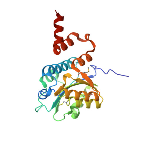
    4WGF, 4WH0

  • PubMed Abstract: 

    As part of the ongoing effort to functionally and structurally characterize virulence factors in the opportunistic pathogen Pseudomonas aeruginosa, we determined the crystal structure of YcaC co-purified with the target protein at resolutions of 2.34 and 2.56 Å without a priori knowledge of the protein identity or experimental phases. The three-dimensional structure of YcaC adopts a well-known cysteine hydrolase fold with the putative active site residues conserved. The active site cysteine is covalently bound to propionamide in one crystal form, whereas the second form contains an S-mercaptocysteine. The precise biological function of YcaC is unknown; however, related prokaryotic proteins have functions in antibacterial resistance, siderophore production and NADH biosynthesis. Here, we show that YcaC is exceptionally well conserved across both bacterial and fungal species despite being non-ubiquitous. This suggests that whilst YcaC may not be part of an integral pathway, the function could confer a significant evolutionary advantage to microbial life.


  • Organizational Affiliation

    School of Biological and Biomedical Sciences, Durham University, Durham DH1 3LE, UK. m.k.groftehauge@durham.ac.uk.


Macromolecules
Find similar proteins by:  (by identity cutoff)  |  3D Structure
Entity ID: 1
MoleculeChains Sequence LengthOrganismDetailsImage
Putative hydrolase
A, B, C, D, E
A, B, C, D, E, F, G, H
205Pseudomonas aeruginosaMutation(s): 0 
UniProt
Find proteins for Q9I4D6 (Pseudomonas aeruginosa (strain ATCC 15692 / DSM 22644 / CIP 104116 / JCM 14847 / LMG 12228 / 1C / PRS 101 / PAO1))
Explore Q9I4D6 
Go to UniProtKB:  Q9I4D6
Entity Groups  
Sequence Clusters30% Identity50% Identity70% Identity90% Identity95% Identity100% Identity
UniProt GroupQ9I4D6
Sequence Annotations
Expand
  • Reference Sequence
Experimental Data & Validation

Experimental Data

  • Method: X-RAY DIFFRACTION
  • Resolution: 2.56 Å
  • R-Value Free: 0.191 
  • R-Value Work: 0.146 
  • R-Value Observed: 0.148 
  • Space Group: P 1 21 1
Unit Cell:
Length ( Å )Angle ( ˚ )
a = 74.46α = 90
b = 114.53β = 91.9
c = 96.16γ = 90
Software Package:
Software NamePurpose
PHENIXrefinement
PHENIXmodel building
PHASERphasing
Aimlessdata scaling
XDSdata reduction

Structure Validation

View Full Validation Report


Entry History & Funding Information

Deposition Data


Funding OrganizationLocationGrant Number
United Kingdom--

Revision History  (Full details and data files)

  • Version 1.0: 2015-07-29
    Type: Initial release
  • Version 1.1: 2024-01-10
    Changes: Data collection, Database references, Derived calculations, Refinement description