4V1F

Crystal structure of a mycobacterial ATP synthase rotor ring in complex with Bedaquiline


Experimental Data Snapshot

  • Method: X-RAY DIFFRACTION
  • Resolution: 1.70 Å
  • R-Value Free: 0.162 
  • R-Value Work: 0.156 
  • R-Value Observed: 0.157 

wwPDB Validation   3D Report Full Report


Ligand Structure Quality Assessment 


This is version 1.4 of the entry. See complete history


Literature

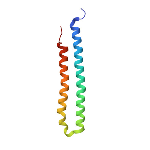
Structure of the mycobacterial ATP synthase Fo rotor ring in complex with the anti-TB drug bedaquiline.

Preiss, L.Langer, J.D.Yildiz, O.Eckhardt-Strelau, L.Guillemont, J.E.Koul, A.Meier, T.

(2015) Sci Adv 1: e1500106-e1500106

  • DOI: https://doi.org/10.1126/sciadv.1500106
  • Primary Citation of Related Structures:  
    4V1F, 4V1G

  • PubMed Abstract: 

    Multidrug-resistant tuberculosis (MDR-TB) is more prevalent today than at any other time in human history. Bedaquiline (BDQ), a novel Mycobacterium-specific adenosine triphosphate (ATP) synthase inhibitor, is the first drug in the last 40 years to be approved for the treatment of MDR-TB. This bactericidal compound targets the membrane-embedded rotor (c-ring) of the mycobacterial ATP synthase, a key metabolic enzyme required for ATP generation. We report the x-ray crystal structures of a mycobacterial c9 ring without and with BDQ bound at 1.55- and 1.7-Å resolution, respectively. The structures and supporting functional assays reveal how BDQ specifically interacts with the rotor ring via numerous interactions and thereby completely covers the c-ring's ion-binding sites. This prevents the rotor ring from acting as an ion shuttle and stalls ATP synthase operation. The structures explain how diarylquinoline chemicals specifically inhibit the mycobacterial ATP synthase and thus enable structure-based drug design of next-generation ATP synthase inhibitors against Mycobacterium tuberculosis and other bacterial pathogens.


  • Organizational Affiliation

    Department of Structural Biology, Max Planck Institute of Biophysics, Max-von-Laue-Straße 3, 60438 Frankfurt am Main, Germany.


Macromolecules
Find similar proteins by:  (by identity cutoff)  |  3D Structure
Entity ID: 1
MoleculeChains Sequence LengthOrganismDetailsImage
F0F1 ATP SYNTHASE SUBUNIT C
A, B, C
86Mycolicibacterium phleiMutation(s): 0 
EC: 3.6.3.14
Entity Groups  
Sequence Clusters30% Identity50% Identity70% Identity90% Identity95% Identity100% Identity
Sequence Annotations
Expand
  • Reference Sequence
Experimental Data & Validation

Experimental Data

  • Method: X-RAY DIFFRACTION
  • Resolution: 1.70 Å
  • R-Value Free: 0.162 
  • R-Value Work: 0.156 
  • R-Value Observed: 0.157 
  • Space Group: H 3
Unit Cell:
Length ( Å )Angle ( ˚ )
a = 75.048α = 90
b = 75.048β = 90
c = 166.55γ = 120
Software Package:
Software NamePurpose
PHENIXrefinement
XDSdata reduction
Aimlessdata scaling
PHASERphasing

Structure Validation

View Full Validation Report


Ligand Structure Quality Assessment 


Entry History 

Deposition Data

Revision History  (Full details and data files)

  • Version 1.0: 2015-05-20
    Type: Initial release
  • Version 1.1: 2015-12-09
    Changes: Database references
  • Version 1.2: 2019-01-16
    Changes: Data collection, Database references
  • Version 1.3: 2020-07-29
    Type: Remediation
    Reason: Carbohydrate remediation
    Changes: Data collection, Derived calculations, Other, Structure summary
  • Version 1.4: 2024-01-10
    Changes: Data collection, Database references, Refinement description, Structure summary