4UM5

Crystal structure of 3-deoxy-D-manno-octulosonate 8-phosphate phosphatase from Moraxella catarrhalis in complex with Magnesium ion and Phosphate ion


Experimental Data Snapshot

  • Method: X-RAY DIFFRACTION
  • Resolution: 2.34 Å
  • R-Value Free: 0.246 
  • R-Value Work: 0.188 
  • R-Value Observed: 0.191 

wwPDB Validation   3D Report Full Report


This is version 1.2 of the entry. See complete history


Literature

Ligand-Bound Structures of 3-Deoxy-D-Manno-Octulosonate 8-Phosphate Phosphatase from Moraxella Catarrhalis Reveal a Water Channel Connecting to the Active Site for the Second Step of Catalysis

Dhindwal, S.Priyadarshini, P.Patil, D.N.Tapas, S.Kumar, P.Tomar, S.Kumar, P.

(2015) Acta Crystallogr D Biol Crystallogr 71: 239

  • DOI: https://doi.org/10.1107/S1399004714025218
  • Primary Citation of Related Structures:  
    4UM5, 4UM7, 4UMD, 4UME, 4UMF

  • PubMed Abstract: 

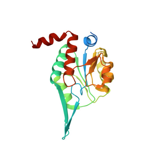
    KdsC, the third enzyme of the 3-deoxy-D-manno-octulosonic acid (KDO) biosynthetic pathway, catalyzes a substrate-specific reaction to hydrolyze 3-deoxy-D-manno-octulosonate 8-phosphate to generate a molecule of KDO and phosphate. KdsC is a phosphatase that belongs to the C0 subfamily of the HAD superfamily. To understand the molecular basis for the substrate specificity of this tetrameric enzyme, the crystal structures of KdsC from Moraxella catarrhalis (Mc-KdsC) with several combinations of ligands, namely metal ion, citrate and products, were determined. Various transition states of the enzyme have been captured in these crystal forms. The ligand-free and ligand-bound crystal forms reveal that the binding of ligands does not cause any specific conformational changes in the active site. However, the electron-density maps clearly showed that the conformation of KDO as a substrate is different from the conformation adopted by KDO when it binds as a cleaved product. Furthermore, structural evidence for the existence of an intersubunit tunnel has been reported for the first time in the C0 subfamily of enzymes. A role for this tunnel in transferring water molecules from the interior of the tetrameric structure to the active-site cleft has been proposed. At the active site, water molecules are required for the formation of a water bridge that participates as a proton shuttle during the second step of the two-step phosphoryl-transfer reaction. In addition, as the KDO biosynthesis pathway is a potential antibacterial target, pharmacophore-based virtual screening was employed to identify inhibitor molecules for the Mc-KdsC enzyme.


  • Organizational Affiliation

    Department of Biotechnology, Indian Institute of Technology Roorkee, Roorkee, Uttarakhand 247 667, India.


Macromolecules
Find similar proteins by:  (by identity cutoff)  |  3D Structure
Entity ID: 1
MoleculeChains Sequence LengthOrganismDetailsImage
3-DEOXY-D-MANNO-OCTULOSONATE 8-PHOSPHATE PHOSPHATASE KDSC
A, B, C, D
193Moraxella catarrhalis BC8Mutation(s): 0 
EC: 3.1.3.45
UniProt
Find proteins for A0A0J9X241 (Moraxella catarrhalis BC8)
Explore A0A0J9X241 
Go to UniProtKB:  A0A0J9X241
Entity Groups  
Sequence Clusters30% Identity50% Identity70% Identity90% Identity95% Identity100% Identity
UniProt GroupA0A0J9X241
Sequence Annotations
Expand
  • Reference Sequence
Small Molecules
Ligands 3 Unique
IDChains Name / Formula / InChI Key2D Diagram3D Interactions
PO4
Query on PO4

Download Ideal Coordinates CCD File 
F [auth A]
G [auth A]
J [auth B]
K [auth B]
M [auth C]
F [auth A],
G [auth A],
J [auth B],
K [auth B],
M [auth C],
N [auth C],
P [auth D],
Q [auth D],
R [auth D]
PHOSPHATE ION
O4 P
NBIIXXVUZAFLBC-UHFFFAOYSA-K
EDO
Query on EDO

Download Ideal Coordinates CCD File 
H [auth A]1,2-ETHANEDIOL
C2 H6 O2
LYCAIKOWRPUZTN-UHFFFAOYSA-N
MG
Query on MG

Download Ideal Coordinates CCD File 
E [auth A],
I [auth B],
L [auth C],
O [auth D]
MAGNESIUM ION
Mg
JLVVSXFLKOJNIY-UHFFFAOYSA-N
Experimental Data & Validation

Experimental Data

  • Method: X-RAY DIFFRACTION
  • Resolution: 2.34 Å
  • R-Value Free: 0.246 
  • R-Value Work: 0.188 
  • R-Value Observed: 0.191 
  • Space Group: P 1 21 1
Unit Cell:
Length ( Å )Angle ( ˚ )
a = 87.795α = 90
b = 45.77β = 90.32
c = 87.801γ = 90
Software Package:
Software NamePurpose
REFMACrefinement
HKL-2000data reduction
SCALEPACKdata scaling
MOLREPphasing

Structure Validation

View Full Validation Report


Entry History 

Deposition Data

Revision History  (Full details and data files)

  • Version 1.0: 2015-02-11
    Type: Initial release
  • Version 1.1: 2015-02-25
    Changes: Database references
  • Version 1.2: 2024-01-10
    Changes: Data collection, Database references, Derived calculations, Other, Refinement description