Molecular basis for the recognition of cyclic-di-AMP by PstA, a PII -like signal transduction protein.
Choi, P.H., Sureka, K., Woodward, J.J., Tong, L.(2015) Microbiologyopen 4: 361-374
- PubMed: 25693966
- DOI: https://doi.org/10.1002/mbo3.243
- Primary Citation of Related Structures:
4RWW, 4RWX - PubMed Abstract:
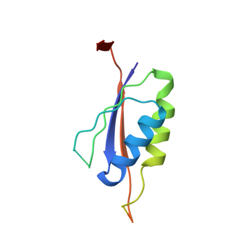
Cyclic-di-AMP (c-di-AMP) is a broadly conserved bacterial second messenger that is of importance in bacterial physiology. The molecular receptors mediating the cellular responses to the c-di-AMP signal are just beginning to be discovered. PstA is a previously uncharacterized PII -like protein which has been identified as a c-di-AMP receptor. PstA is widely distributed and conserved among Gram-positive bacteria in the phylum Firmicutes. Here, we report the biochemical, structural, and functional characterization of PstA from Listeria monocytogenes. We have determined the crystal structures of PstA in the c-di-AMP-bound and apo forms at 1.6 and 2.9 Å resolution, respectively, which provide the molecular basis for its specific recognition of c-di-AMP. PstA forms a homotrimer structure that has overall similarity to the PII protein family which binds ATP. However, PstA is markedly different from PII proteins in the loop regions, and these structural differences mediate the specific recognition of their respective nucleotide ligand. The residues composing the c-di-AMP binding pocket are conserved, suggesting that c-di-AMP recognition by PstA is of functional importance. Disruption of pstA in L. monocytogenes affected c-di-AMP-mediated alterations in bacterial growth and lysis. Overall, we have defined the PstA family as a conserved and specific c-di-AMP receptor in bacteria.
Organizational Affiliation:
Department of Biological Sciences, Columbia University, New York City, New York, 10027.