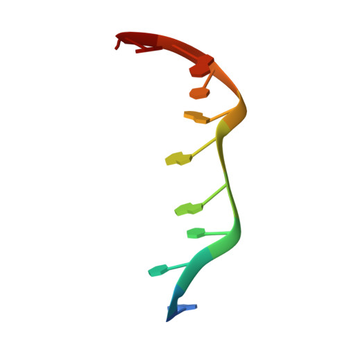
An intercalation-locked parallel-stranded DNA tetraplex.
Tripathi, S., Zhang, D., Paukstelis, P.J.(2015) Nucleic Acids Res 43: 1937-1944
- PubMed: 25628357
- DOI: https://doi.org/10.1093/nar/gkv033
- Primary Citation of Related Structures:
4RIM, 4RIP - PubMed Abstract:
DNA has proved to be an excellent material for nanoscale construction because complementary DNA duplexes are programmable and structurally predictable. However, in the absence of Watson-Crick pairings, DNA can be structurally more diverse. Here, we describe the crystal structures of d(ACTCGGATGAT) and the brominated derivative, d(AC(Br)UCGGA(Br)UGAT). These oligonucleotides form parallel-stranded duplexes with a crystallographically equivalent strand, resulting in the first examples of DNA crystal structures that contains four different symmetric homo base pairs. Two of the parallel-stranded duplexes are coaxially stacked in opposite directions and locked together to form a tetraplex through intercalation of the 5'-most A-A base pairs between adjacent G-G pairs in the partner duplex. The intercalation region is a new type of DNA tertiary structural motif with similarities to the i-motif. (1)H-(1)H nuclear magnetic resonance and native gel electrophoresis confirmed the formation of a parallel-stranded duplex in solution. Finally, we modified specific nucleotide positions and added d(GAY) motifs to oligonucleotides and were readily able to obtain similar crystals. This suggests that this parallel-stranded DNA structure may be useful in the rational design of DNA crystals and nanostructures.
Organizational Affiliation:
Department of Chemistry & Biochemistry, Center for Biomolecular Structure & Organization, Maryland Nanocenter, University of Maryland, College Park, MD 20742, USA.