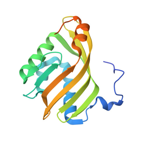
X-ray crystallographic validation of structure predictions used in computational design for protein stabilization.
Floor, R.J., Wijma, H.J., Jekel, P.A., Terwisscha van Scheltinga, A.C., Dijkstra, B.W., Janssen, D.B.(2015) Proteins 83: 940-951
- PubMed: 25739581
- DOI: https://doi.org/10.1002/prot.24791
- Primary Citation of Related Structures:
4R9K, 4R9L - PubMed Abstract:
Protein engineering aimed at enhancing enzyme stability is increasingly supported by computational methods for calculation of mutant folding energies and for the design of disulfide bonds. To examine the accuracy of mutant structure predictions underlying these computational methods, crystal structures of thermostable limonene epoxide hydrolase variants obtained by computational library design were determined. Four different predicted effects indeed contributed to the obtained stabilization: (i) enhanced interactions between a flexible loop close to the N-terminus and the rest of the protein; (ii) improved interactions at the dimer interface; (iii) removal of unsatisfied hydrogen bonding groups; and (iv) introduction of additional positively charged groups at the surface. The structures of an eightfold and an elevenfold mutant showed that most mutations introduced the intended stabilizing interactions, and side-chain conformations were correctly predicted for 72-88% of the point mutations. However, mutations that introduced a disulfide bond in a flexible region had a larger influence on the backbone conformation than predicted. The enzyme active sites were unaltered, in agreement with the observed preservation of catalytic activities. The structures also revealed how a c-Myc tag, which was introduced for facile detection and purification, can reduce access to the active site and thereby lower the catalytic activity. Finally, sequence analysis showed that comprehensive mutant energy calculations discovered stabilizing mutations that are not proposed by the consensus or B-FIT methods.
Organizational Affiliation:
Biotransformation and Biocatalysis, Groningen Biomolecular Sciences and Biotechnology Institute, University of Groningen, Nijenborgh 4, 9747 AG, Groningen, The Netherlands.