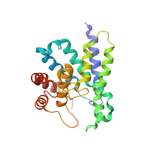
Structural basis of functional diversification of the HD-GYP domain revealed by the Pseudomonas aeruginosa PA4781 protein, which displays an unselective bimetallic binding site.
Rinaldo, S., Paiardini, A., Stelitano, V., Brunotti, P., Cervoni, L., Fernicola, S., Protano, C., Vitali, M., Cutruzzola, F., Giardina, G.(2015) J Bacteriol 197: 1525-1535
- PubMed: 25691523
- DOI: https://doi.org/10.1128/JB.02606-14
- Primary Citation of Related Structures:
4R8Z - PubMed Abstract:
The intracellular level of the bacterial secondary messenger cyclic di-3',5'-GMP (c-di-GMP) is determined by a balance between its biosynthesis and degradation, the latter achieved via dedicated phosphodiesterases (PDEs) bearing a characteristic EAL or HD-GYP domain. We here report the crystal structure of PA4781, one of the three Pseudomonas aeruginosa HD-GYP proteins, which we have previously characterized in vitro. The structure shows a bimetallic active site whose metal binding mode is different from those of both HD-GYP PDEs characterized so far. Purified PA4781 does not contain iron in the active site as for other HD-GYPs, and we show that it binds to a wide range of transition metals with similar affinities. Moreover, the structural features of PA4781 indicate that this is preferentially a pGpG binding protein, as we previously suggested. Our results point out that the structural features of HD-GYPs are more complex than predicted so far and identify the HD-GYP domain as a conserved scaffold which has evolved to preferentially interact with a partner GGDEF but which harbors different functions obtained through diversification of the active site. In bacteria, the capability to form biofilms, responsible for increased pathogenicity and antibiotic resistance, is almost universally stimulated by the second messenger cyclic di-GMP (c-di-GMP). To design successful strategies for targeting biofilm formation, a detailed characterization of the enzymes involved in c-di-GMP metabolism is crucial. We solved the structure of the HD-GYP domain of PA4781 from Pseudomonas aeruginosa, involved in c-di-GMP degradation. This is the third structure of this class of phosphodiesterases to be solved, and with respect to its homologues, it shows significant differences both in the nature and in the binding mode of the coordinated metals, indicating that HD-GYP proteins are able to fine-tune their function, thereby increasing the chances of the microorganism to adapt to different environmental needs.
Organizational Affiliation:
Istituto Pasteur-Fondazione Cenci Bolognetti, Department of Biochemical Sciences, Sapienza University of Rome, Rome, Italy.


