4QUV

Structure of an integral membrane delta(14)-sterol reductase


Experimental Data Snapshot

  • Method: X-RAY DIFFRACTION
  • Resolution: 2.74 Å
  • R-Value Free: 0.284 
  • R-Value Work: 0.233 
  • R-Value Observed: 0.235 

wwPDB Validation   3D Report Full Report


Ligand Structure Quality Assessment 


This is version 1.3 of the entry. See complete history


Literature

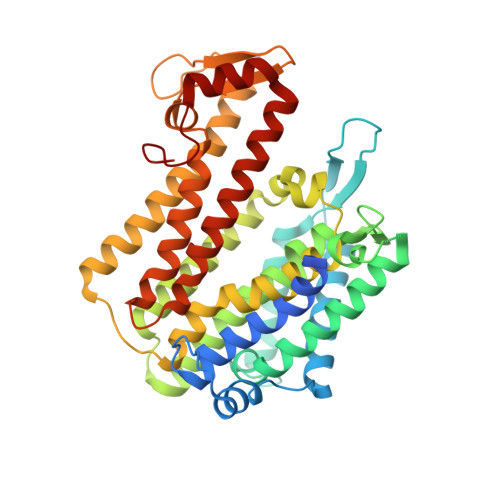
Structure of an integral membrane sterol reductase from Methylomicrobium alcaliphilum.

Li, X.Roberti, R.Blobel, G.

(2015) Nature 517: 104-107

  • DOI: https://doi.org/10.1038/nature13797
  • Primary Citation of Related Structures:  
    4QUV

  • PubMed Abstract: 

    Sterols are essential biological molecules in the majority of life forms. Sterol reductases including Δ(14)-sterol reductase (C14SR, also known as TM7SF2), 7-dehydrocholesterol reductase (DHCR7) and 24-dehydrocholesterol reductase (DHCR24) reduce specific carbon-carbon double bonds of the sterol moiety using a reducing cofactor during sterol biosynthesis. Lamin B receptor (LBR), an integral inner nuclear membrane protein, also contains a functional C14SR domain. Here we report the crystal structure of a Δ(14)-sterol reductase (MaSR1) from the methanotrophic bacterium Methylomicrobium alcaliphilum 20Z (a homologue of human C14SR, LBR and DHCR7) with the cofactor NADPH. The enzyme contains ten transmembrane segments (TM1-10). Its catalytic domain comprises the carboxy-terminal half (containing TM6-10) and envelops two interconnected pockets, one of which faces the cytoplasm and houses NADPH, while the other one is accessible from the lipid bilayer. Comparison with a soluble steroid 5β-reductase structure suggests that the reducing end of NADPH meets the sterol substrate at the juncture of the two pockets. A sterol reductase activity assay proves that MaSR1 can reduce the double bond of a cholesterol biosynthetic intermediate, demonstrating functional conservation to human C14SR. Therefore, our structure as a prototype of integral membrane sterol reductases provides molecular insight into mutations in DHCR7 and LBR for inborn human diseases.


  • Organizational Affiliation

    Laboratory of Cell Biology, Howard Hughes Medical Institute, The Rockefeller University, New York, New York 10065, USA.


Macromolecules
Find similar proteins by:  (by identity cutoff)  |  3D Structure
Entity ID: 1
MoleculeChains Sequence LengthOrganismDetailsImage
Delta(14)-sterol reductase
A, B
427Methylotuvimicrobium alcaliphilum 20ZMutation(s): 0 
Gene Names: ergMEALZ_1312
EC: 1.3.1.70
Membrane Entity: Yes 
UniProt
Find proteins for G4SW86 (Methylotuvimicrobium alcaliphilum (strain DSM 19304 / NCIMB 14124 / VKM B-2133 / 20Z))
Explore G4SW86 
Go to UniProtKB:  G4SW86
Entity Groups  
Sequence Clusters30% Identity50% Identity70% Identity90% Identity95% Identity100% Identity
UniProt GroupG4SW86
Sequence Annotations
Expand
  • Reference Sequence
Small Molecules
Ligands 1 Unique
IDChains Name / Formula / InChI Key2D Diagram3D Interactions
NDP
Query on NDP

Download Ideal Coordinates CCD File 
C [auth A],
D [auth B]
NADPH DIHYDRO-NICOTINAMIDE-ADENINE-DINUCLEOTIDE PHOSPHATE
C21 H30 N7 O17 P3
ACFIXJIJDZMPPO-NNYOXOHSSA-N
Experimental Data & Validation

Experimental Data

  • Method: X-RAY DIFFRACTION
  • Resolution: 2.74 Å
  • R-Value Free: 0.284 
  • R-Value Work: 0.233 
  • R-Value Observed: 0.235 
  • Space Group: P 1
Unit Cell:
Length ( Å )Angle ( ˚ )
a = 74.66α = 66
b = 74.615β = 90.37
c = 79.549γ = 86.86
Software Package:
Software NamePurpose
HKL-2000data collection
SOLVEphasing
PHENIXrefinement
HKL-2000data reduction
HKL-2000data scaling

Structure Validation

View Full Validation Report


Ligand Structure Quality Assessment 


Entry History 

Deposition Data

Revision History  (Full details and data files)

  • Version 1.0: 2014-10-08
    Type: Initial release
  • Version 1.1: 2014-10-29
    Changes: Database references
  • Version 1.2: 2015-01-28
    Changes: Database references
  • Version 1.3: 2024-02-28
    Changes: Data collection, Database references, Derived calculations