4QJ8

Crystal structure of inactive HIV-1 protease variant (I50V/A71V) in complex with p1-p6 substrate variant (P453L)


Experimental Data Snapshot

  • Method: X-RAY DIFFRACTION
  • Resolution: 2.00 Å
  • R-Value Free: 0.239 
  • R-Value Work: 0.172 
  • R-Value Observed: 0.175 

wwPDB Validation   3D Report Full Report


This is version 1.3 of the entry. See complete history


Literature

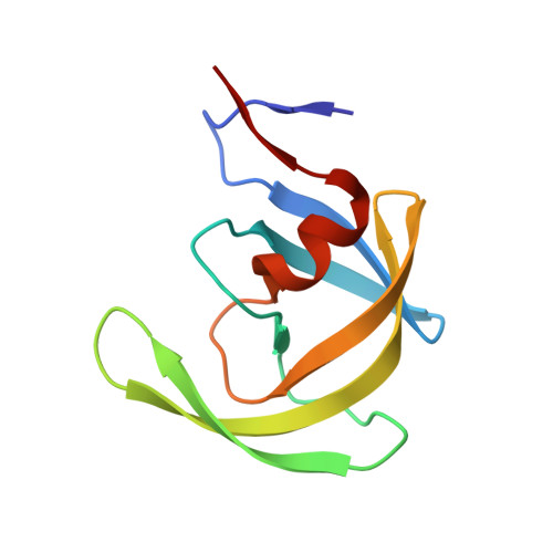
Structural basis and distal effects of Gag substrate coevolution in drug resistance to HIV-1 protease.

Ozen, A.Lin, K.H.Kurt Yilmaz, N.Schiffer, C.A.

(2014) Proc Natl Acad Sci U S A 111: 15993-15998

  • DOI: https://doi.org/10.1073/pnas.1414063111
  • Primary Citation of Related Structures:  
    4QJ2, 4QJ6, 4QJ7, 4QJ8, 4QJ9, 4QJA

  • PubMed Abstract: 

    Drug resistance mutations in response to HIV-1 protease inhibitors are selected not only in the drug target but elsewhere in the viral genome, especially at the protease cleavage sites in the precursor protein Gag. To understand the molecular basis of this protease-substrate coevolution, we solved the crystal structures of drug resistant I50V/A71V HIV-1 protease with p1-p6 substrates bearing coevolved mutations. Analyses of the protease-substrate interactions reveal that compensatory coevolved mutations in the substrate do not restore interactions lost due to protease mutations, but instead establish other interactions that are not restricted to the site of mutation. Mutation of a substrate residue has distal effects on other residues' interactions as well, including through the induction of a conformational change in the protease. Additionally, molecular dynamics simulations suggest that restoration of active site dynamics is an additional constraint in the selection of coevolved mutations. Hence, protease-substrate coevolution permits mutational, structural, and dynamic changes via molecular mechanisms that involve distal effects contributing to drug resistance.


  • Organizational Affiliation

    Department of Biochemistry and Molecular Pharmacology, University of Massachusetts Medical School, Worcester, MA 01605.


Macromolecules
Find similar proteins by:  (by identity cutoff)  |  3D Structure
Entity ID: 1
MoleculeChains Sequence LengthOrganismDetailsImage
Protease
A, B, C, D
99HIV-1 M:B_ARV2/SF2Mutation(s): 5 
Gene Names: gag-pol
EC: 3.4.23.16
UniProt
Find proteins for P03369 (Human immunodeficiency virus type 1 group M subtype B (isolate ARV2/SF2))
Explore P03369 
Go to UniProtKB:  P03369
Entity Groups  
Sequence Clusters30% Identity50% Identity70% Identity90% Identity95% Identity100% Identity
UniProt GroupP03369
Sequence Annotations
Expand
  • Reference Sequence

Find similar proteins by:  Sequence   |   3D Structure  

Entity ID: 2
MoleculeChains Sequence LengthOrganismDetailsImage
p1-p6 peptide
E, F
10HIV-1 M:B_ARV2/SF2Mutation(s): 0 
UniProt
Find proteins for P03349 (Human immunodeficiency virus type 1 group M subtype B (isolate ARV2/SF2))
Explore P03349 
Go to UniProtKB:  P03349
Entity Groups  
Sequence Clusters30% Identity50% Identity70% Identity90% Identity95% Identity100% Identity
UniProt GroupP03349
Sequence Annotations
Expand
  • Reference Sequence
Experimental Data & Validation

Experimental Data

  • Method: X-RAY DIFFRACTION
  • Resolution: 2.00 Å
  • R-Value Free: 0.239 
  • R-Value Work: 0.172 
  • R-Value Observed: 0.175 
  • Space Group: P 1 21 1
Unit Cell:
Length ( Å )Angle ( ˚ )
a = 51.306α = 90
b = 62.663β = 97.97
c = 57.477γ = 90
Software Package:
Software NamePurpose
HKL-2000data collection
PHASERphasing
REFMACrefinement
HKL-2000data reduction
HKL-2000data scaling

Structure Validation

View Full Validation Report


Entry History 

Deposition Data

Revision History  (Full details and data files)

  • Version 1.0: 2014-10-29
    Type: Initial release
  • Version 1.1: 2014-11-26
    Changes: Database references
  • Version 1.2: 2014-12-03
    Changes: Database references
  • Version 1.3: 2024-02-28
    Changes: Data collection, Database references, Derived calculations