4P4B

X-ray structure of human glutamate carboxypeptidase II (GCPII) in complex with a phosphoramidate inhibitor CTT54


Experimental Data Snapshot

  • Method: X-RAY DIFFRACTION
  • Resolution: 1.93 Å
  • R-Value Free: 0.178 
  • R-Value Work: 0.155 
  • R-Value Observed: 0.156 

wwPDB Validation   3D Report Full Report


Ligand Structure Quality Assessment 


This is version 2.0 of the entry. See complete history


Literature

Design of composite inhibitors targeting glutamate carboxypeptidase II: the importance of effector functionalities.

Novakova, Z.Cerny, J.Choy, C.J.Nedrow, J.R.Choi, J.K.Lubkowski, J.Berkman, C.E.Barinka, C.

(2016) FEBS J 283: 130-143

  • DOI: https://doi.org/10.1111/febs.13557
  • Primary Citation of Related Structures:  
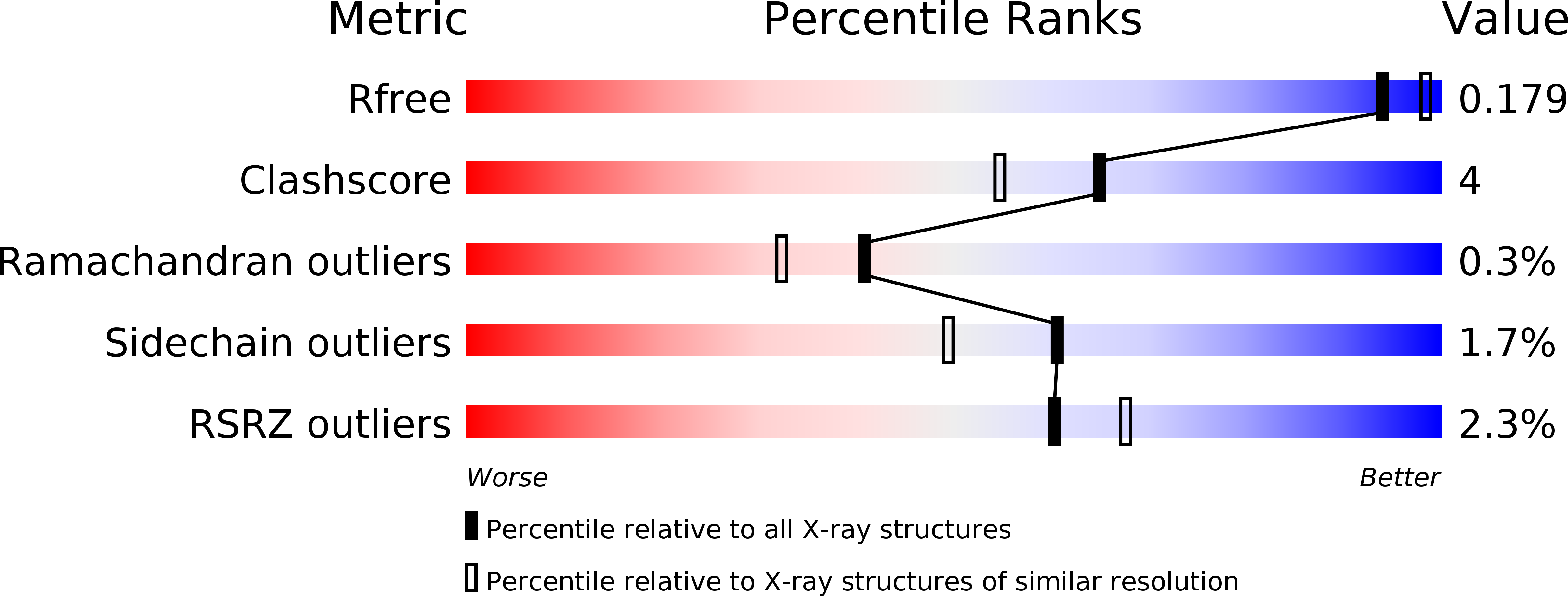
    4P44, 4P45, 4P4B, 4P4D, 4P4E, 4P4F, 4P4I, 4P4J

  • PubMed Abstract: 

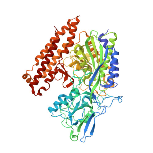
    Inhibitors targeting human glutamate carboxypeptidase II (GCPII) typically consist of a P1' glutamate-derived binding module, which warrants the high affinity and specificity, linked to an effector function that is positioned within the entrance funnel of the enzyme. Here we present a comprehensive structural and computational study aimed at dissecting the importance of the effector function for GCPII binding and affinity. To this end we determined crystal structures of human GCPII in complex with a series of phosphoramidate-based inhibitors harboring effector functions of diverse physicochemical characteristics. Our data show that higher binding affinities of phosphoramidates, compared to matching phosphonates, are linked to the presence of additional hydrogen bonds between Glu424 and Gly518 of the enzyme and the amide group of the phosphoramidate. While the positioning of the P1' glutamate-derived module within the S1' pocket of GCPII is invariant, interaction interfaces between effector functions and residues lining the entrance funnel are highly varied, with the positively charged arginine patch defined by Arg463, Arg534 and Arg536 being the only 'hot-spot' common to several studied complexes. This variability stems in part from the fact that the effector/GCPII interfaces generally encompass isolated areas of nonpolar residues within the entrance funnel and resulting van der Waals contacts lack the directionality typical for hydrogen bonding interactions. The presented data unravel a complexity of binding modes of inhibitors within non-prime site(s) of GCPII and can be exploited for the design of novel GCPII-specific compounds.


  • Organizational Affiliation

    Institute of Biotechnology, Academy of Sciences of the Czech Republic, Prague, Czech Republic.


Macromolecules
Find similar proteins by:  (by identity cutoff)  |  3D Structure
Entity ID: 1
MoleculeChains Sequence LengthOrganismDetailsImage
Glutamate carboxypeptidase 2707Homo sapiensMutation(s): 0 
Gene Names: FOLH1FOLHNAALAD1PSMPSMAGIG27
EC: 3.4.17.21
UniProt & NIH Common Fund Data Resources
Find proteins for Q04609 (Homo sapiens)
Explore Q04609 
Go to UniProtKB:  Q04609
PHAROS:  Q04609
GTEx:  ENSG00000086205 
Entity Groups  
Sequence Clusters30% Identity50% Identity70% Identity90% Identity95% Identity100% Identity
UniProt GroupQ04609
Sequence Annotations
Expand
  • Reference Sequence
Oligosaccharides

Help

Entity ID: 2
MoleculeChains Length2D Diagram Glycosylation3D Interactions
2-acetamido-2-deoxy-beta-D-glucopyranose-(1-4)-2-acetamido-2-deoxy-beta-D-glucopyranose
B, C, D
2N-Glycosylation
Glycosylation Resources
GlyTouCan:  G42666HT
GlyCosmos:  G42666HT
GlyGen:  G42666HT
Entity ID: 3
MoleculeChains Length2D Diagram Glycosylation3D Interactions
beta-D-mannopyranose-(1-4)-2-acetamido-2-deoxy-beta-D-glucopyranose-(1-4)-2-acetamido-2-deoxy-beta-D-glucopyranose
E
3N-Glycosylation
Glycosylation Resources
GlyTouCan:  G15407YE
GlyCosmos:  G15407YE
GlyGen:  G15407YE
Entity ID: 4
MoleculeChains Length2D Diagram Glycosylation3D Interactions
alpha-D-mannopyranose-(1-3)-beta-D-mannopyranose-(1-4)-2-acetamido-2-deoxy-beta-D-glucopyranose-(1-4)-2-acetamido-2-deoxy-beta-D-glucopyranose
F
4N-Glycosylation
Glycosylation Resources
GlyTouCan:  G81315DD
GlyCosmos:  G81315DD
GlyGen:  G81315DD
Small Molecules
Ligands 5 Unique
IDChains Name / Formula / InChI Key2D Diagram3D Interactions
2G2
Query on 2G2

Download Ideal Coordinates CCD File 
M [auth A]L-gamma-glutamyl-O-[(S)-{[(1S)-1,3-dicarboxypropyl]amino}(hydroxy)phosphoryl]-L-serine
C13 H22 N3 O12 P
KSAFNYJPIDGLBS-FXQIFTODSA-N
NAG
Query on NAG

Download Ideal Coordinates CCD File 
K [auth A],
L [auth A]
2-acetamido-2-deoxy-beta-D-glucopyranose
C8 H15 N O6
OVRNDRQMDRJTHS-FMDGEEDCSA-N
ZN
Query on ZN

Download Ideal Coordinates CCD File 
G [auth A],
H [auth A]
ZINC ION
Zn
PTFCDOFLOPIGGS-UHFFFAOYSA-N
CA
Query on CA

Download Ideal Coordinates CCD File 
I [auth A]CALCIUM ION
Ca
BHPQYMZQTOCNFJ-UHFFFAOYSA-N
CL
Query on CL

Download Ideal Coordinates CCD File 
J [auth A]CHLORIDE ION
Cl
VEXZGXHMUGYJMC-UHFFFAOYSA-M
Binding Affinity Annotations 
IDSourceBinding Affinity
2G2 BindingDB:  4P4B IC50: 14 (nM) from 1 assay(s)
Binding MOAD:  4P4B IC50: 14 (nM) from 1 assay(s)
Experimental Data & Validation

Experimental Data

  • Method: X-RAY DIFFRACTION
  • Resolution: 1.93 Å
  • R-Value Free: 0.178 
  • R-Value Work: 0.155 
  • R-Value Observed: 0.156 
  • Space Group: I 2 2 2
Unit Cell:
Length ( Å )Angle ( ˚ )
a = 101.634α = 90
b = 130.307β = 90
c = 158.577γ = 90
Software Package:
Software NamePurpose
REFMACrefinement
HKL-2000data reduction
HKL-2000data scaling

Structure Validation

View Full Validation Report


Ligand Structure Quality Assessment 


Entry History 

Deposition Data

  • Released Date: 2015-05-20 
  • Deposition Author(s): Barinka, C.

Revision History  (Full details and data files)

  • Version 1.0: 2015-05-20
    Type: Initial release
  • Version 1.1: 2015-11-04
    Changes: Database references
  • Version 1.2: 2016-01-20
    Changes: Database references
  • Version 2.0: 2020-07-29
    Type: Remediation
    Reason: Carbohydrate remediation
    Changes: Atomic model, Data collection, Derived calculations, Structure summary