4OLS

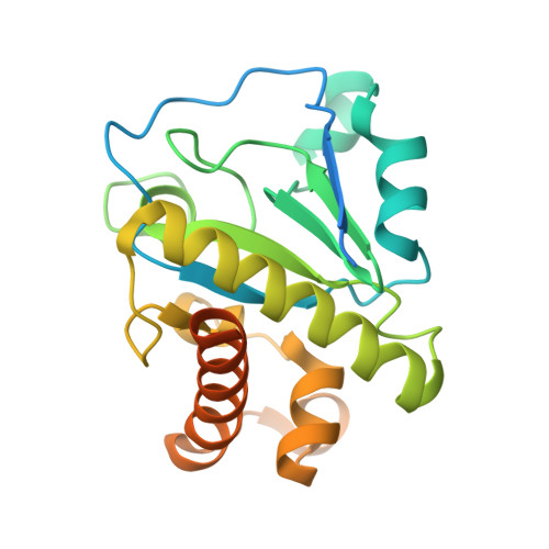
The amidase-2 domain of LysGH15


Experimental Data Snapshot

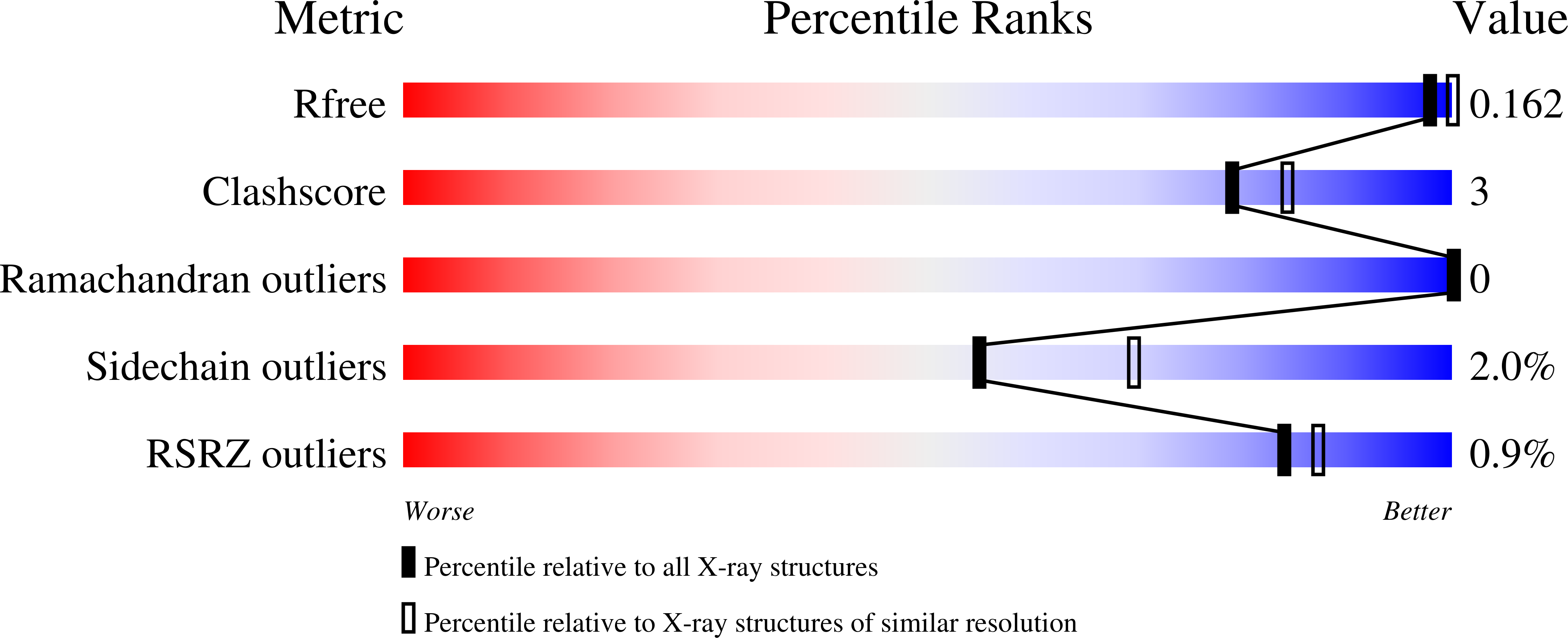
  • Method: X-RAY DIFFRACTION
  • Resolution: 2.27 Å
  • R-Value Free: 0.161 
  • R-Value Work: 0.158 
  • R-Value Observed: 0.158 

wwPDB Validation   3D Report Full Report


This is version 1.2 of the entry. See complete history


Literature

Structural and biochemical characterization reveals LysGH15 as an unprecedented "EF-hand-like" calcium-binding phage lysin.

Gu, J.Feng, Y.Feng, X.Sun, C.Lei, L.Ding, W.Niu, F.Jiao, L.Yang, M.Li, Y.Liu, X.Song, J.Cui, Z.Han, D.Du, C.Yang, Y.Ouyang, S.Liu, Z.J.Han, W.

(2014) PLoS Pathog 10: e1004109-e1004109

  • DOI: https://doi.org/10.1371/journal.ppat.1004109
  • Primary Citation of Related Structures:  
    2MK5, 4OLK, 4OLS

  • PubMed Abstract: 

    The lysin LysGH15, which is derived from the staphylococcal phage GH15, demonstrates a wide lytic spectrum and strong lytic activity against methicillin-resistant Staphylococcus aureus (MRSA). Here, we find that the lytic activity of the full-length LysGH15 and its CHAP domain is dependent on calcium ions. To elucidate the molecular mechanism, the structures of three individual domains of LysGH15 were determined. Unexpectedly, the crystal structure of the LysGH15 CHAP domain reveals an "EF-hand-like" calcium-binding site near the Cys-His-Glu-Asn quartet active site groove. To date, the calcium-binding site in the LysGH15 CHAP domain is unique among homologous proteins, and it represents the first reported calcium-binding site in the CHAP family. More importantly, the calcium ion plays an important role as a switch that modulates the CHAP domain between the active and inactive states. Structure-guided mutagenesis of the amidase-2 domain reveals that both the zinc ion and E282 are required in catalysis and enable us to propose a catalytic mechanism. Nuclear magnetic resonance (NMR) spectroscopy and titration-guided mutagenesis identify residues (e.g., N404, Y406, G407, and T408) in the SH3b domain that are involved in the interactions with the substrate. To the best of our knowledge, our results constitute the first structural information on the biochemical features of a staphylococcal phage lysin and represent a pivotal step forward in understanding this type of lysin.


  • Organizational Affiliation

    Key Laboratory of Zoonosis, Ministry of Education, College of Veterinary Medicine, Jilin University, Changchun, China.


Macromolecules
Find similar proteins by:  (by identity cutoff)  |  3D Structure
Entity ID: 1
MoleculeChains Sequence LengthOrganismDetailsImage
Endolysin
A, B, C, D
242Staphylococcus phage G15Mutation(s): 0 
Gene Names: lysGH15GH15_071
UniProt
Find proteins for D6QY02 (Staphylococcus phage G15)
Explore D6QY02 
Go to UniProtKB:  D6QY02
Entity Groups  
Sequence Clusters30% Identity50% Identity70% Identity90% Identity95% Identity100% Identity
UniProt GroupD6QY02
Sequence Annotations
Expand
  • Reference Sequence
Small Molecules
Ligands 3 Unique
IDChains Name / Formula / InChI Key2D Diagram3D Interactions
ZN
Query on ZN

Download Ideal Coordinates CCD File 
E [auth A]
F [auth A]
G [auth A]
H [auth A]
I [auth B]
E [auth A],
F [auth A],
G [auth A],
H [auth A],
I [auth B],
J [auth B],
K [auth B],
L [auth B],
M [auth B],
N [auth C],
O [auth C],
P [auth C],
Q [auth C],
R [auth C],
S [auth D],
T [auth D],
U [auth D]
ZINC ION
Zn
PTFCDOFLOPIGGS-UHFFFAOYSA-N
FE
Query on FE

Download Ideal Coordinates CCD File 
W [auth D]FE (III) ION
Fe
VTLYFUHAOXGGBS-UHFFFAOYSA-N
MG
Query on MG

Download Ideal Coordinates CCD File 
V [auth D]MAGNESIUM ION
Mg
JLVVSXFLKOJNIY-UHFFFAOYSA-N
Experimental Data & Validation

Experimental Data

  • Method: X-RAY DIFFRACTION
  • Resolution: 2.27 Å
  • R-Value Free: 0.161 
  • R-Value Work: 0.158 
  • R-Value Observed: 0.158 
  • Space Group: P 63
Unit Cell:
Length ( Å )Angle ( ˚ )
a = 135.4α = 90
b = 135.4β = 90
c = 107.077γ = 120
Software Package:
Software NamePurpose
HKL-2000data collection
SHELXSphasing
REFMACrefinement
HKL-2000data reduction
HKL-2000data scaling

Structure Validation

View Full Validation Report


Entry History 

Deposition Data

Revision History  (Full details and data files)

  • Version 1.0: 2014-06-04
    Type: Initial release
  • Version 1.1: 2019-12-18
    Changes: Database references
  • Version 1.2: 2024-03-20
    Changes: Data collection, Database references, Derived calculations